Conjugation of a low affinity nerve growth factor receptor (p75NTR) antibody to saporin has produced a cytotoxin that eliminates the CBF neurons, while sparing neighboring neurons that express GAD, calbindin and parvalbumin. mu p75-SAP eliminates cells expressing p75NTR in mouse. Permanent and selective removal of cholinergic forebrain neurons makes an important animal model for the study of behavior, neuronal loss (e.g. Alzheimer’s disease), plasticity of other systems in response to loss, replacement therapy, and drug effects and dependence.
mu p75-SAP is a chemical conjugate of the affinity-purified rabbit polyclonal antibody p75NTR and the ribosome-inactivating protein, saporin. It specifically eliminates p75NTR-positive cells in mouse.
To eliminate p75NTR-expressing cells in rat, use 192-IgG-SAP (Cat. #IT-01). To eliminate p75NTR-expressing cells in other mammals, use ME20.4-SAP (Cat. #IT-15).
mu p75-SAP is available individually (Cat. #IT-16) or as a kit (Cat. #KIT-16) which includes mu p75-SAP and Rabbit IgG-SAP (Cat. #IT-35).
keywords: P75NTR, NGFR, Alzheimer’s Disease, dementia, CBF neurons, cholinergic forebrain, animal model, neuronal loss, low affinity nerve growth factor, neurotrophin receptors, mesenchyme, Anti-NGFR, Anti-Nerve Growth Factor, mup75, mu p75, cholinergic forebrain neurons, Upper cervical ganglion, brain, neuroscience
Minocycline protects basal forebrain cholinergic neurons from mu p75-saporin immunotoxic lesioning.
Hunter CL, Quintero EM, Gilstrap L, Bhat NR, Granholm AC (2004) Minocycline protects basal forebrain cholinergic neurons from mu p75-saporin immunotoxic lesioning. Eur J Neurosci 19(12):3305-3316. doi: 10.1111/j.0953-816X.2004.03439.x
Summary: In Alzheimer’s disease basal cholinergic degeneration is accompanied by glial activation and the release of pro-inflammatory cytokines. To investigate whether neural events other than degeneration can cause effects of Alzheimer’s disease, the authors treated mice with minocycline after lesioning the basal forebrain with 3.6 µg of mu p75-SAP (Cat. #IT-16). Administration of minocycline reduced the loss of cholinergic neurons, reduced glial response to the lesion, and lessened the cognitive impairment due to mu p75-SAP lesions.
Related Products: mu p75-SAP (Cat. #IT-16)
Selective immunolesions of cholinergic neurons in mice: effects on neuroanatomy, neurochemistry, and behavior.
Berger-Sweeney JE, Stearns NA, Murg SL, Floerke-Nashner LR, Lappi DA, Baxter MG (2001) Selective immunolesions of cholinergic neurons in mice: effects on neuroanatomy, neurochemistry, and behavior. J Neurosci 21(20):8164-8173. doi: 10.1523/JNEUROSCI.21-20-08164.2001
Summary: 192-Saporin (Cat. #IT-01) has long been an effective agent for elimination of cholinergic neurons in the basal forebrain of rats. Until the development of mu p75-SAP (Cat. #IT-16) there was no equivalent agent for use in mice. The authors tested mu p75-SAP in vitro and in vivo (1.8-3.6 µg in right lateral ventricle), using cytotoxic, histochemical, and behavioral assays. The data shows that mu p75-SAP is a highly selective and efficacious lesioning agent for cholinergic neurons in the mouse. The authors conclude that mu p75-SAP will be a powerful tool to use in combination with genetic modification to investigate cholinergic damage in mouse models of Alzheimer’s disease.
Related Products: mu p75-SAP (Cat. #IT-16), 192-IgG-SAP (Cat. #IT-01)
Cholinergic basal forebrain lesion decreases neurotrophin signaling without affecting tau hyperphosphorylation in genetically susceptible mice.
Turnbull M, Coulson E (2017) Cholinergic basal forebrain lesion decreases neurotrophin signaling without affecting tau hyperphosphorylation in genetically susceptible mice. J Alzheimers Dis 55:1141-1154.. doi: 10.3233/JAD-160805
Summary: Alzheimer’s disease(AD) is a progressive, irreversible neurodegenerative disease that destroys memory and cognitive function. Aggregates of hyperphosphorylated tau protein are a prominent feature in the brain of patients with AD, and area major contributor to neuronal toxicity and disease progression. However, the factors that initiate the toxic cascade that results in tau hyperphosphorylation in AD are unknown. The authors investigated whether degeneration of basal forebrain cholinergic neurons (BFCNs) and/or resultant decrease in neurotrophin signaling cause aberrant tau hyperphosphorylation. Two-month-old male and female pR5 mice were infused with murine p75-SAP (Cat. #IT-16) at a concentration of 0.4 mg/ml or 0.4 mg/ml of control Rabbit IgG-SAP (Cat. #IT-35) using a 30G needle attached to a 5 ml Hamilton syringe and pump. The needle was lowered into the medial septum according to coordinates in a mouse brain atlas, and the toxin was infused at a rate of 0.4 ul/min (1.5 u total volume). The results reveal that the loss of BFCNs in pre-symptomatic pR5 tau transgenic mice results in a decrease in hippocampal brain-derived neurotrophic factor levels and reduced TrkB receptor activation. However, there was no exacerbation of the levels of phosphorylated tau or its aggregation in the hippocampus of susceptible mice. Furthermore the animals’ performance in a hippocampal-dependent learning and memory task was unaltered, and no changes in hippocampal synaptic markers were observed. This suggests that tau pathology is likely to be regulated independently of BFCN degeneration and the corresponding decrease in hippocampal neurotrophin levels, although these features may still contribute to disease etiology.
Related Products: mu p75-SAP (Cat. #IT-16), Rabbit IgG-SAP (Cat. #IT-35)
Saporin as a commercial reagent: its uses and unexpected impacts in the biological sciences-tools from the plant kingdom
Ancheta LR, Shramm PA, Bouajram R, Higgins D, Lappi DA (2022) Saporin as a commercial reagent: its uses and unexpected impacts in the biological sciences-tools from the plant kingdom. Toxins (Basel) 14(3):184. doi: 10.3390/toxins14030184 PMID: 35324681
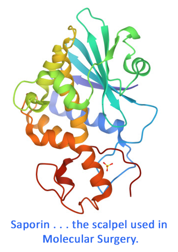
Summary: Saporin is a ribosome-inactivating protein that can cause inhibition of protein synthesis and causes cell death when delivered inside a cell. Development of commercial Saporin results in a technology termed ‘molecular surgery’, with Saporin as the scalpel. Its low toxicity (it has no efficient method of cell entry) and sturdy structure make Saporin a safe and simple molecule for many purposes. The most popular applications use experimental molecules that deliver Saporin via an add-on targeting molecule. These add-ons come in several forms: peptides, protein ligands, antibodies, even DNA fragments that mimic cell-binding ligands. Cells that do not express the targeted cell surface marker will not be affected. This review will highlight some newer efforts and discuss significant and unexpected impacts on science that molecular surgery has yielded over the last almost four decades. There are remarkable changes in fields such as the Neurosciences with models for Alzheimer’s Disease and epilepsy, and game-changing effects in the study of pain and itch. Many other uses are also discussed to record the wide-reaching impact of Saporin in research and drug development.
Read complete article.
browse all references for this product | back to top


Reviews
There are no reviews yet.