Targeting in mouse: mu p75-SAP (Cat. #IT-16)
To create this targeted toxin, we affinity-purified the rabbit polyclonal with the immunogen bound to a solid support, and conjugated the affinity-purified antibody (Cat. #AB-N01AP) to saporin. As can be seen in the cytotoxicity assay below, mu p75-SAP has an EC50 in the picomolar range. This greater potency translates to smaller amounts used for elimination of p75-positive neurons in the mouse brain, and results in a greater index of efficacy and lesser non-specific cytotoxicity.
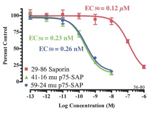
NG3 cells are plated at 1000 cells/well and incubated overnight. Saporin and mu p75-SAP are added in 10-μl volumes and the plates are incubated 72 hours. PMS/MTS developing reagent is added and the plates are incubated 1-2 hours, then read at 490 nm.
Targeting in rat: 192-IgG-SAP (Cat. #IT-01)
Intraventricular injection of 192- IgG-SAP (192-Saporin) results in almost complete elimination of LNGFR (p75NTR)-positive cells in rat. 192-IgG- SAP is directed to a cell-surface antigen that is only expressed at high levels on neurons in the cholinergic basal forebrain (CBF). The antigen, p75NTR, is not expressed on the neighboring, non-cholinergic neurons. Visit our website to browse through the more than 370 scientific publications with this powerful targeting tool.
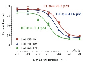
7H6 cells are plated at 1000 cells/well and incubated overnight. 192 IgG-SAP lots are added in 10-μl volumes and the plates are incubated 72 hours. PMS/MTS developing reagent is added and the plates are incubated 0.5 to 1 hour, then read at 490 nm.
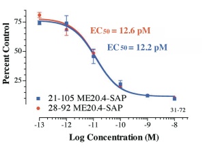
Targeting in other species: ME20.4-SAP (Cat. #IT-15)
This immunotoxin provides researchers with a powerful lesioning tool — more specific and effective than chemical, surgical or electrolytic lesioning and is active in several species (rabbit, sheep, dog, cat, raccoon, pig and several primate species). Intraventricular injection of ME20.4-SAP has been used to eliminate low-affinity nerve growth factor receptor (p75NTR)-positive cells. Tissue-directed injection has also been used in primates to cause loss of p75NTR-positive neurons.