Intraventricular injection of 192-IgG-SAP (192-Saporin) results in almost complete elimination of LNGFR (p75NTR)-positive cells in rat. 192-IgG-SAP is directed to a cell-surface antigen that is only expressed at high levels on neurons in the cholinergic basal forebrain (CBF). The antigen, p75NTR, is not expressed on the neighboring, non-cholinergic neurons. 192-IgG-SAP specifically eliminates cholinergic neurons of the basal forebrain, medial septum, diagonal band of Broca, nucleus basalis of Meynert, and Purkinje neurons of the cerebellum. It provides researchers with a powerful lesioning tool – more specific and effective than chemical, surgical or electrolytic lesioning. Permanent and selective removal of cholinergic forebrain neurons makes an important animal model for the study of behavior, neuronal loss (e.g. Alzheimer’s disease), plasticity of other systems in response to loss, replacement therapy, and drug effects and dependence.
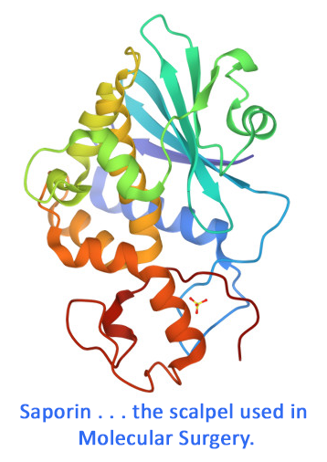
192-IgG-SAP is a chemical conjugate of the mouse monoclonal antibody to rat p75NTR and the ribosome-inactivating protein, saporin. It specifically eliminates cells expressing p75NTR, also known as the low affinity nerve growth factor receptor.
To eliminate p75NTR-expressing cells in mouse, use mu p75-SAP (Cat. #IT-16). To eliminate p75NTR-expressing cells in other mammals, use ME20.4-SAP (Cat. #IT-15). See also NGFR (192-IgG, p75) Mouse Monoclonal (Cat. #AB-N43).
192-IgG-SAP is available individually (Cat. #IT-01) or as a kit (Cat. #KIT-01) which includes 192-IgG-SAP and Mouse IgG-SAP (Cat. #IT-18).
keywords: P75NTR, NGFR, Alzheimer’s Disease, dementia, basal forebrain, animal model, neuronal loss, low affinity nerve growth factor, neurotrophin receptors, mesenchyme, Anti-NGFR, Anti-Nerve Growth Factor, 192-IgG, p75, cholinergic forebrain neurons, Upper cervical ganglion, rat, CBF neurons, brain, neuroscience
Selective immunolesions of cholinergic neurons in mice: effects on neuroanatomy, neurochemistry, and behavior.
Berger-Sweeney JE, Stearns NA, Murg SL, Floerke-Nashner LR, Lappi DA, Baxter MG (2001) Selective immunolesions of cholinergic neurons in mice: effects on neuroanatomy, neurochemistry, and behavior. J Neurosci 21(20):8164-8173. doi: 10.1523/JNEUROSCI.21-20-08164.2001
Summary: 192-Saporin (Cat. #IT-01) has long been an effective agent for elimination of cholinergic neurons in the basal forebrain of rats. Until the development of mu p75-SAP (Cat. #IT-16) there was no equivalent agent for use in mice. The authors tested mu p75-SAP in vitro and in vivo (1.8-3.6 µg in right lateral ventricle), using cytotoxic, histochemical, and behavioral assays. The data shows that mu p75-SAP is a highly selective and efficacious lesioning agent for cholinergic neurons in the mouse. The authors conclude that mu p75-SAP will be a powerful tool to use in combination with genetic modification to investigate cholinergic damage in mouse models of Alzheimer’s disease.
Related Products: mu p75-SAP (Cat. #IT-16), 192-IgG-SAP (Cat. #IT-01)
Pain sensitivity following loss of cholinergic basal forebrain (CBF) neurons in the rat.
Vierck C, Yezierski R, Wiley R (2016) Pain sensitivity following loss of cholinergic basal forebrain (CBF) neurons in the rat. Neuroscience 319:23-34. doi: 10.1016/j.neuroscience.2016.01.038
Objective: There is a large amount of research on the involvement of cholinergic mechanisms on spinal transmission of pain signals, indicating that cholinergic agonists can attenuate this kind of pain. In contrast, some studies have shown affective reactions to pain are suppressed by cholinergic antagonists. The authors investigated the disagreement between reflexive and affective reactions.
Summary: Lesioned rats displayed decreased escape from thermal stimulation, as well as loss of the normal hyperalgesic effect of sound stress. Results indicate that the basal forebrain cholinergic system plays a role in central processing of pain.
Usage: Administration of 192-IgG-SAP with a 4-μg injection into the left lateral ventricle of rats. Animals were tested in temperature escape and sound stress models.
Related Products: 192-IgG-SAP (Cat. #IT-01)
Cannabinoid receptors and glial response following a basal forebrain cholinergic lesion
Llorente-Ovejero A, Bengoetxea de Tena I, Martínez-Gardeazabal J, Moreno-Rodríguez M, Lombardero L, Manuel I, Rodríguez-Puertas R (2022) Cannabinoid receptors and glial response following a basal forebrain cholinergic lesion. ACS Pharmacol Transl Sci 5(9):791-802. doi: 10.1021/acsptsci.2c00069 PMID: 36110372
Objective: The endocannabinoid system is involved in the control of learning, memory, and neuroinflammatory processes and plays a role in neurodegeneration, such as in Alzheimer’s disease (AD). The objective was to study the roles of cannabinoid receptors in the regulation of neuroinflammation.
Summary: Selective agonists and antagonists to the cannabinoid receptors CB1 and CB2 were studied for their binding to G-proteins after specific lesioning of the basal forebrain cholinergic neurons (BCFN) using 192-IgG-SAP. These neurons are the same cholinergic pathways that are lost in the early stages of Alzheimer's disease (AD). In their study, an increase of microglia immunoreactivities (GFAP and Iba-1) and decrease of astrocyte immunoreactivities were seen which indicate microglia-mediated neuroinflammation. In cortical BFCN projection areas, CB1 receptor binding to Gi/o-proteins was upregulated and at the injection site, the area that showed the highest increase of microglia, only slight CB2 binding to Gi/o-proteins were detected. Dose: Rats received 135 ng/μLof 192IgG-saporin (1μL/hemisphere; 0.25μL/min).
Related Products: 192-IgG-SAP (Cat. #IT-01)
Neurotoxic effects, mechanisms, and outcome of 192 IgG-Saporin lesions.
Petrosini L, De Bartolo P, Cutuli D (2021) Neurotoxic effects, mechanisms, and outcome of 192 IgG-Saporin lesions. RM Kostrzewa (Ed.): Handbook of Neurotoxicity . Springer, Cham doi: 10.1007/978-3-030-71519-9_79-1
Summary: 192-IgG-saporin selectively destroys basal forebrain cholinergic neurons that provide cholinergic input to the hippocampus, entire cortical mantle, amygdala, and olfactory bulb. Immunotoxic lesions by 192-IgG-saporin represent a valid animal model of Alzheimer’s disease, given the degeneration of basal cholinergic system present in this pathology. The selective lesioning of cholinergic innervation by means of 192-IgG-saporin (injected i.p. or i.c.v.) is able to interfere with experience-dependent plasticity.
Related Products: 192-IgG-SAP (Cat. #IT-01)
browse all references for this product | back to top


Reviews
There are no reviews yet.