Today we’re highlighting one of our saporin conjugates, Anti-CD117-SAP. This is a tool for depleting cells that express CD117, the tyrosine kinase growth factor receptor.
CD117 is expressed on hematopoietic stem cells, mast cells, and acute myeloid leukemia cells. Some of the areas where CD117 plays an essential role is in the regulation of cell survival and proliferation, hematopoiesis, stem cell maintenance and mast cell development, migration and function.
Allogeneic hematopoietic stem cell transplantation can cure genetic diseases such as severe combined immunodeficiency (SCID), but it’s associated with significant toxicities such as graft-versus-host disease (GvHD). Using a patients’ own gene-modified hematopoietic stem and progenitor cells (HSPCs) can eliminate the risk of GvHD, but the treatment relies on gene transfer using viruses and genotoxic conditioning which carries risks.
The authors describe how base-editing is an alternative means to correct genetic defects while engineered virus-like particles (eVLPs) can deliver the base-edited proteins without the risks seen with viral integration.
In conclusion, transplantation of HSPCs that have been repaired through eVLP-mediated base-editing with CD117-ADC conditioning successfully reversed the SCID phenotype in mice, and highlights a significant advancement in disease treatment without the current toxicities.
We are highlighting one of our saporin conjugates, Neurotensin-SAP — a tool for depleting cells that express the neurotensin receptor (NTSR).
Neurotensin is a 13-amino-acid peptide found in the brain and spinal cord and is released by the hypothalamus. It is synthesized as part of a larger precursor protein that also includes the related neuropeptide neuromedin N. Neurotensin affects pituitary hormone release, interacts with the dopaminergic system, and is involved in vasodilation and hypotension. It can also modulate pain perception where intrathecal neurotensin has also been shown to be anti-nociceptive.
The objective was to determine whether NTSR1-expressing enteropancreatic neurons mediate the glucose-lowering effects of dietary olive oil and neurotensin, and to characterize their physiological role in glucose homeostasis.
The authors unilaterally injected neurotensin-SAP into the nodose ganglia to ablate NTSR1-expressing vagal neurons.
In their study, they demonstrated that neurotensin improves glucose tolerance by activating NTSR1-expressing enteropancreatic neurons, which connect the gut and pancreas. Ablation or disruption of these neurons abolished the glucoregulatory effects of both neurotensin and olive oil, establishing their necessity and sufficiency in this pathway.
Kozlíková M, Aukrust IKF, Rohlíčková M, Macháček M, Berg K, Weyergang A, Selbo PK (2026) Photochemical enhancement of PD-L1-SAP immunotoxin efficacy in non-small cell lung cancer cell lines. Front Immunol 17:1750003. doi: 10.3389/fimmu.2026.1750003 PMID: 41909645
Objective: To investigate photochemical internalization (PCI), a light–controlled endosomal escape technology, as a strategy to enhance intracellular delivery and efficacy of a PD–L1–targeted immunotoxin (anti–PD–L1–SAP).
Summary: Non–small cell lung cancer (NSCLC) cell lines with high (NCI–H1975) and low (A549) PD–L1 expression were subjected to PCI, resulting in a pronounced increase in cytotoxicity with picomolar potency (30 pM), while A549 cells required a higher dose (1000 pM) for a similar effect. PCI enhances delivery and activity of PD–L1–targeted biologics and may help overcome resistance mechanisms. Overall, PCI expands the therapeutic window of PD–L1–targeted immunotoxins and may complement current immunotherapies, supporting further preclinical evaluation in NSCLC.
Usage: Cells were then incubated with TPCS2a and anti-PD-L1-SAP (IT-45) for 18 h. The concentration of anti-PD-L1-SAP was 30, 100 and 1000 pM for NCI-H1975 cell line and 1000 pM for A549 cell line. BIgG-SAP (IT-74) was used as control.
Chen J, Xu T, Zhang C, Li L, He Y, Sun Z, He J, Yao Z, Cai P, Huang Y, Ye F, Guo W, Jia M, Qu J, Chen JF, Zhang Y (2026) 40 Hz light flickering alleviates chronic pain via adenosine signaling in the retina-amygdala pathway. Cell Res doi: 10.1038/s41422-026-01227-7 PMID: 41781500
Objective: To demonstrate the multifaceted therapeutic benefits of 40 Hz light flickering as a novel non-invasive approach for pain management and reveal a distinct retina-central amygdala circuit and adenosine signaling mechanism for control of chronic pain and pain memory
Summary: The authors examined the role of intrinsically photosensitive RGCs (ipRGCs). To selectively ablate ipRGCs, melanopsin-SAP was bilaterally injected into the vitreous humor. Treated mice displayed no significant change in baseline paw withdrawal threshold. Importantly, the antinociceptive effects of 40 Hz light flickering remained intact in both CFA and SNI models, despite ablation of ipRGCs. These results indicate that ipRGCs are not essential for the analgesia induced by 40 Hz light flickering.
Usage: Melanopsin-SAP (IT-44)) was bilaterally injected into the vitreous humor.
Wang W, Zhang Y, Li L, Xu Y, Zhang W, Chen X, Wang X, Tong G, Zhang P (2026) PACAP/PAC 1 modulates light-induced sleep via the ipRGC-VLPO pathway. Biochem Biophys Res Commun 808:153462. doi: 10.1016/j.bbrc.2026.153462 PMID: 41702189
Objective: To investigate the mechanism of PACAP in ipRGC–VLPO light-induced sleep.
Summary: Partial ablation of ipRGCs by Melanopsin-SAP reduced light-induced sleep duration, whereas PACAP 1-38 administration reversed this effect, leading to an increase in REM sleep. After the partial destruction of ipRGCs through the intraocular injection of saporin (SAP), we continued to observe the effect of PACAP on light-induced sleep. The results showed that after the microdialysis injection of PACAP 1-38 into the VLPO of SAP mice, the light-induced sleep of the mice increased; specifically, REM sleep significantly increased. The results suggest that PACAP is involved in ipRGC–VLPO-mediated light-induced sleep.
Maria NI, Xia Y, Raparia C, Lin K, Martinez S, Yi Z, Zhang W, Aziz M, Wang P, Guerra M, Salmon, J, Sones JL, Arazi A, Hoover P, Davidson A (2026) Pregnancy loss due to early developmental defects in lupus mice expressing human TLR8. bioRxiv 2026.02.07.701591. doi: 10.64898/2026.02.07.701591
Objective: To explore human TLR8 (huTLR8) in anti-phospholipid (APL) antibody-mediated placental injury.
Summary: The authors used a novel mouse model of pregnancy loss in SLE-prone mice expressing huTLR8. Findings indicate huTLR8, likely through myeloid cell activation and cytolytic T cell recruitment, drives placental injury in the context of SLE and APL autoantibodies.
Usage: 8- to 10-week-old Sle1.CD45.1 mice were treated with biotin anti-CD117 and streptavidin-saporin
Gao JL, Li Z, Calderon-Perez R, Pavek A, Kim L, McDermott DH, Murphy PM (2026) Gene therapy via CRISPR/Cas9-mediated Cxcr4 disease allele inactivation reverses leukopenia in WHIM mice. J Clin Invest 136(5):e202073. doi: 10.1172/JCI202073 PMID: 41505207
Objective: To provide proof of principle that CRISPR/Cas9-mediated inactivation of the Cxcr4 disease allele, combined with nongenotoxic HSC- targeted conditioning, may offer a safe and effective gene therapy strategy generalizable to all WHIM-causing mutations.
Summary: Warts, hypogammaglobulinemia, infections, and myelokathexis (WHIM) syndrome is an immunodeficiency caused by autosomal dominant hyperfunctional mutations in chemokine receptor CXCR4 that promote panleukopenia due to BM retention. The authors modified their previous protocol by adding conditioning with a nongenotoxic CD117-targeted immunotoxin, CD117-antibody-saporin-conjugate.
Gupta S (2026) Modelling Temporal Lobe Epilepsy with Sclerosis in the SSP-Saporin ‘Trojan Horse’ Model. Univ Calgary Thesis.
Objective: To confirm that the SSP-SAP model reproduces the defining features of human TLE and that a primary and selective GABAergic defect is sufficient to trigger epileptogenesis that results in TLE-HS+.
Summary: The author hypothesized that intrahippocampal SSP-SAP administration will first cause rats to exhibit reactive behavioural seizures, followed by self-generated epileptiform activity that will develop into electrographic seizures. Rats will also develop TLE anatomically characterized by progressive principal cell loss and astrogliosis.
Usage: Rats were stereotaxically injected with 150 nL of SSP-SAP (IT-11, 0.04 ng/nL) at 4 sites in the hippocampus.
McCallum S, Suresh KB, Islam TS, Tripathi MK, Saustad AW, Shelest O, Patil A, Lee D, Kwon B, Leitholf K, Yenokian I, Shaka SE, Beveridge CH, Manchandra P, Randolph CE, Meares GP, Dutta R, Plummer J, Calsavara VF, Kawaguchi R, Knott SRV, Chopra G, Burda JE (2026) Lesion-remote astrocytes govern microglia-mediated white matter repair. Nature 649(8098):959-970. doi: 10.1038/s41586-025-09887-y PMID: 41407858
Objective: To investigate Lesion-remote astrocytes (LRAs) from spared regions of mouse spinal cord following traumatic spinal cord injury.
Summary: The nature of astrocyte-extrinsic mechanisms that trigger discrete reactivity states are not well understood. Using Ccn1 expression as a biomarker of a molecularly distinct white matter LRA reactivity state, the authors explored the mechanism of its induction. IB4-SAP and CTB-SAP were injected in mice to selectively degenerate myelinated or non-myelinated sensory afferents, respectively.
Usage: 8 μg (10μl of 0.8 μg μl−1 in PBS) of Saporin (PR-01), IB4-SAP (IT-10) or CTB-SAP (IT-14) was injected subcutaneously into the plantar surface of the left hindpaw foot pad using a 30G insulin syringe.
Today’s topic is time-course for our saporin conjugates. Common questions we get are (1) how long does it takes to see cell death, (2) how long should I wait before performing histology on an animal, or (3) how long before I see behavioral changes in an animal?
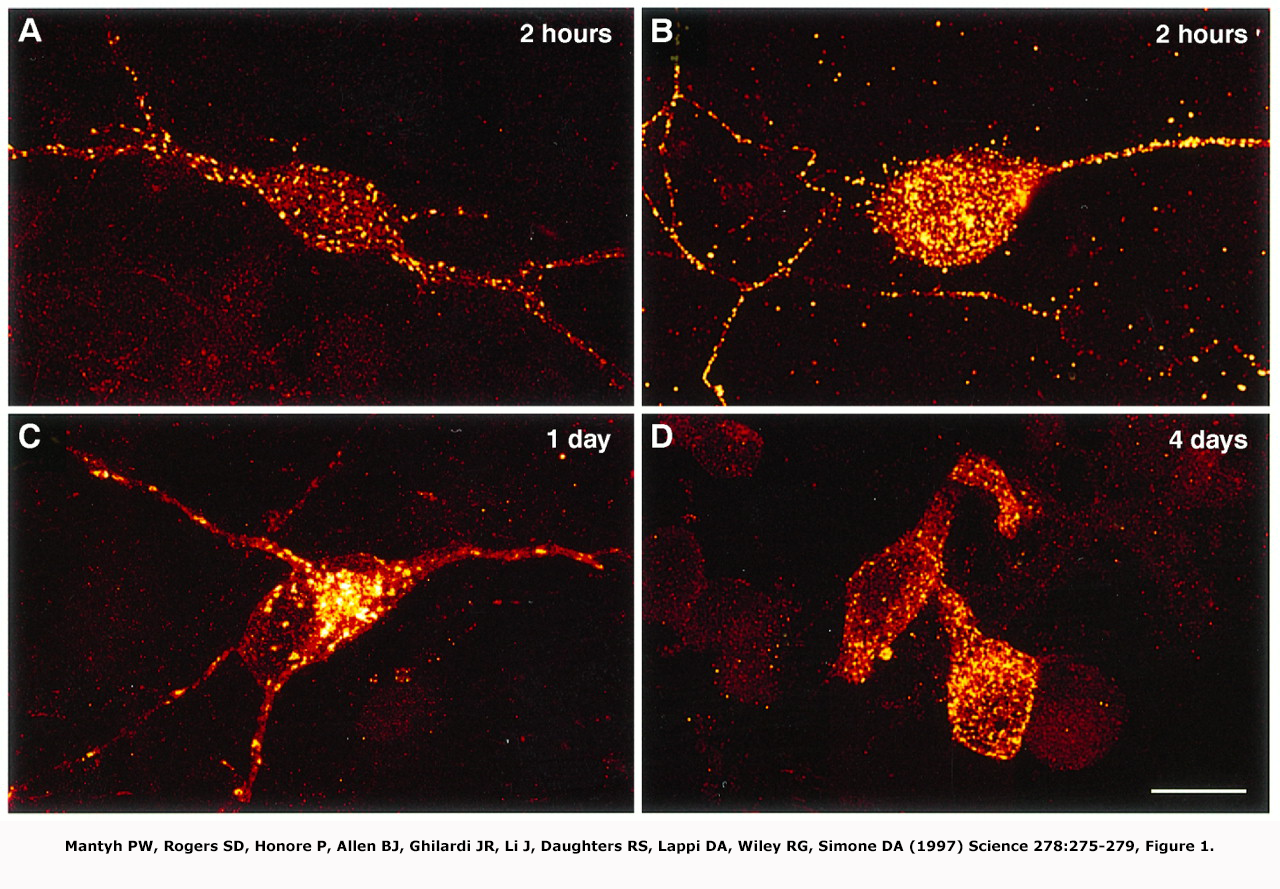
In this image from Mantyh et al. (1997), we are looking at confocal imagery of the binding and internalization of our peptide conjugate SP-SAP to the NK1r receptor in primary cultures of neonatal spinal cord neurons.
As you can see, conjugate binding occurs immediately where within hours, the SP-SAP has recognized and bound to the NK1 receptor. Here we see the areas of concentrated NK1R expression marked by yellow immunofluorescence.
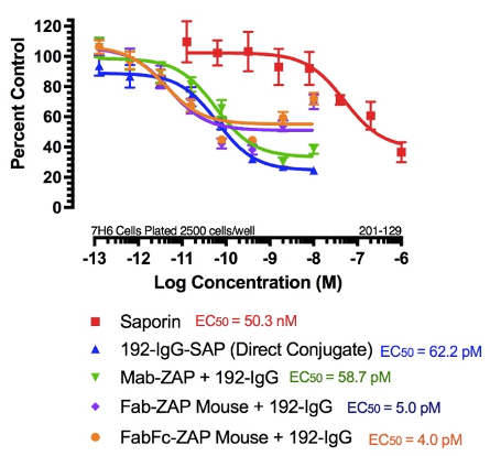
But how long until you see cell death? Here is a cytotoxicity graph of in vitro data of our antibody conjugate 192-IgG-SAP. These are typical data after cells have been treated for 3 days, which is standard protocol.
Waite et al. (1994) used 192-IgG-SAP and showed the appearance of behavioral effects associated with neuronal loss at day four and plateauing at day 7.
This coincides with the time course seen in vitro. At this point, microglia will infiltrate, but stop at 7 days, which is probably the peak day for infiltration. Once there is complete removal of the detritus, microglia down-regulate and at 14 days all debris is cleared and the animal begins to regain normal eating and sleeping habits. This is the idea behind waiting about 2 weeks before performing histology.