We have been using your human fab-pHast (PH-01) product for a while now and are very happy with the results in our imaging applications (we have seen signal to noise of over 70k). However I have recently tried running a black bottom plate on a plate reader, same set up, and am getting a high background signal. My signal to noise is about 1.5 in the best case scenario. Could you please advise how to increase the signal to noise in the plate reader format using fab-pHast?
Answer:
We recommend washing the cells with PBS before reading on the plate reader to help reduce background signal. You can find an internalization assay protocol here.
Question:
I’m using complete media (phenol red DMEM with 10% FBS) for my experiments, could this be contributing to background signal?
Answer:
Using phenol red-containing media could contribute to background signal. Replacing the media with PBS should reduce background significantly.
Question:
Do you have any recommendations on whether to use black opaque-bottom plates, black clear-bottom plates, or standard clear TC plates for maximal S/N?
Answer:
Clear-bottom plates offer the advantage of imaging, but they may introduce more background noise. If imaging is a priority, you might need to balance between signal strength and versatility.
Related Products
All of our pHast Conjugates are available with these fluorescent dyes and as a multi-color kit with all four colors in one in one bundle.
The pHast BLUE has an excitation wavelength of 362 nm with an emission maxima at 452 nm. (PH-B)
The pHast GREEN version has an excitation wavelength of 453 nm with an emission maxima at 522 nm. (PH-G)
The pHast RED has an excitation wavelength of 532 nm with an emission maxima at 560 nm. (PH-R)
The pHast FAR-RED has an excitation wavelength of 643 nm with an emission maxima at 660 nm. (PH-F)
Hello, I am very interested in using your Fab-ZAP line. So Fab-ZAPs simply tells us what mAbs internalize best – is that right? Can you send me some example data? I’d love to see a cell line that has been evaluated and the data that has been generated.
Answer:
Yes, Fab-ZAPs will tell you what mAbs internalize best. We have a variety of different versions of these products (ZAP secondary conjugates) which you can find here.
These have been tested on many different cell lines, both in-house and by our customers.
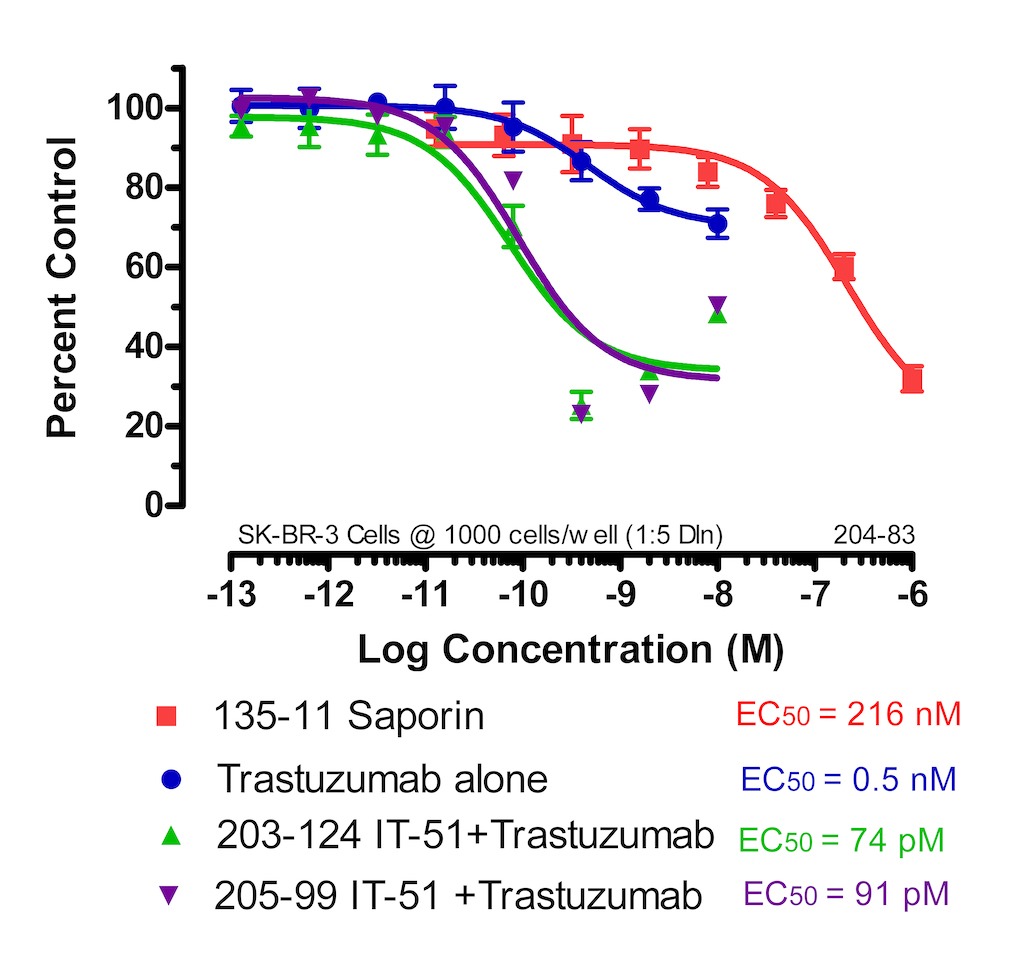
I’ve included a graph that corresponds to Fab-ZAP human (IT-51). Here is a cytotox which tests Fab-ZAP human with Trastuzumab on SK-BR-3 cells (breast cancer cell line).
SK-BR-3 cells were plated at 1000 cells/90 μl/well and incubated overnight. Trastuzumab and Saporin dilutions were made in cell media and 10 μl was added to each well. The Trastuzumab was also diluted in cell media containing, at a final concentration, 4.5 nM/10 μl Fab-ZAP, and 10 μl was added to each well. The plates were incubated for 72 hours. The plates were developed using a solution of XTT/PMS and read at 450 nm. Cytotoxicity was analyzed by comparing well readings of the treated wells to those of the control wells, expressed as a percentage. The number of viable cells remaining on the day of development is measured via cell metabolism of a colorimetric molecule within the developing reagents. Analysis was performed using Prism software (GraphPad, San Diego).
Related Products
ZAP conjugates – These are non-targeted saporin conjugates that “piggyback” on to your primary targeting agent (biotinylated material or antibody) to eliminate specific cells and reveal cell function.
Where does the saporin payload release after internalization? For example, does it require trafficking into a late endosome/lysosomal compartment?
Answer:
Thank you for reaching out to us. Hopefully I can help answer some of your questions regarding what happens to saporin after being internalized.
I would first like to refer you to an article we published, titled “Streptavidin-Saporin: Converting Biotinylated Materials into Targeted Toxins”. In it we review the internalization of saporin and include a few references for support. To answer your question in general, yes the conjugate is typically endocytosed and makes its way to the late endosome.
As an overview of this debated topic, the Wensley, H.J. et al 2019 article (ref #4) studied the escape of saporin from the late endosome and examined the endocytic process to quantify the endosomal escape into the cytosol. The Holmes, S.E et al 2015 (ref #5) and Giansanti, F. et al 2018 (ref #6) articles describe additional research examining chemical and genetic strategies used in assisting in saporin’s escape from the endosome. After endocytocis, Vago, R. et al 2005 (ref #7) compared saporin and ricin A chain and other bacterial toxins looking at their different intracellular routes to enter the cytosol. These articles should provide a nice foundation and hopefully better answer any questions.
If you’re interested in visualizing lysosomal trafficking, you might consider our pHast product line. These are secondary pH-dependent fluorescent conjugates, meaning that they only fluoresce once inside the endosomes and lysosomes of cells (which are acidic compared to the cytosol).
Related Products
pHast Conjugates – one of our pHastest tools for quantitative testing.
Question: I was wondering if you could elaborate on why the Streptavidin-ZAP product recommends to be used at an equimolar ratio with the targeting reagent, when it is capable of binding up to four biotins/molecule?
Answer: It’s a question we get asked sometimes and it’s a good question.
You are correct that streptavidin is capable of binding up to 4 biotin molecules. However, when we created streptavidin-ZAP with the purpose of being a modular way of creating targeted toxins, we learned that the best general rule to follow was using a equimolar reaction. In theory, it is a 1:1 ratio of targeting molecule to streptavidin-ZAP, where we are most likely seeing an average of 1:1, but there is also the possibility of mixed ratios.
The amount of publications using the equimolar approach gave the desired results whether they were using a small biotinylated peptide or whole IgG. You’ll notice that depending on the MW of your biotinylated targeting agent, the amount of streptavidin-ZAP needed for the experiment can vary drastically and through in-house characterization, the equimolar approach still worked best.
Another reason we recommend a 1:1 ratio is based on our experience with our other secondary conjugates. It may be intuitive to think that using a higher dose of targeting agent would induce more cell death, but we found the opposite effect, where the excess, un-reacted targeting agent competed with the conjugated material for surface binding sites, which in turn decreased the amount of saporin being delivered. We have a publication (PMCID: PMC8952126 ) that also describes this observation.
Once you’ve created a baseline using the equimolar protocol and are more accustomed to how streptavidin-ZAP works in your application, please contact us if you feel more optimization is needed. It will be easier to help trouble-shoot when we are all working off the same protocol.
Q: I read on your website that, “There are two types of RIPs: type I, which are much less cytotoxic due to the lack of the B chain and type II, which are distinguished from type I RIPs by the presence of the B chain and their ability to enter cells on their own.”
In the IT-27 Streptavidin-ZAP product, which type of saporin is there? Is it both type I and type II because the saporin is purified from the plant, or is it one specific type only in the product.
A: All saporin molecules are Type I ribosome-inactivating proteins. We only use saporin. An example of a Type II RIP is ricin, which can enter a cell on its own and has been used throughout history as a method of assassination.
Streptavidin-ZAP is streptavidin attached to saporin. On its own it has no way to get inside a cell. By mixing Streptavidin-ZAP with a biotinylated molecule that is recognized on the cell surface, the resulting conjugate is able to bind and internalize saporin into a cell. Once inside saporin inactivates the ribosomes which causes cell death.
Q: When using any of your Fab-Zap product line, the recommended final concentration is 4.5 nM. Is this based on experiments you have done? I question if at 4.5 nM my primary antibody will be saturated with Fab-ZAP secondary conjugate?
A: Yes, the 4.5 nM concentration is what we use to quality-control test our Fab-ZAP conjugates and why we recommend it in the literature. We also recommend only titrating your primary antibody. The 4.5 nM of Fab-ZAP should be enough to saturate your primary antibody. If you have a test of ~10 nM of primary antibody and you experience less cell death than ~1 nM, this will indicate “antibody competition” (i.e., your primary antibody is not saturated). The data sheet shows a cytotox with a nice example of this. (Fab-ZAP data sheet)
Q: What are neuropeptide-toxins and how do they work?
A: Neuropeptide-toxin conjugates are made up of the ribosome-inactivating protein, saporin, coupled to a naturally-occurring or synthetically-modified neuropeptide such as Substance P or dermorphin. The conjugate has binding specificity similar to the native, unconjugated neuropeptide. When the neuropeptide binds to its cognate receptor, the conjugate is internalized. Once inside the target cell within an endosome, the neuropeptide and saporin separate and some of the saporin translocates into the cytoplasm where it catalytically inactivates ribosomes resulting in cell death.
Q: Are neuropeptide-toxins effective suicide transport agents?
A: The general answer to this question is not currently known. However, in the instance of intrathecally injected dermorphin-SAP (Cat. #IT-12), the evidence does NOT favor suicide transport of the neuropeptide-toxin conjugate. When supramaximal doses of dermorphin-SAP (750 ng) are injected into the lumbar subarachnoid space of adult rats, less than 1% of lumbar dorsal root ganglion cells show evidence of saporin activity. This is in spite of the fact that many of these neurons express the targeted mu opioid receptor on their central terminals in the superficial dorsal horn of the spinal cord. This assertion is based on analysis of over 16,000 neurons from dorsal root ganglia in six rats.
Q: Does Mab-ZAP (Cat. #IT-04) bind to the FC portion of mouse IgG?
A: The antibody used to create our Mab-ZAP (IT-04), will react with whole molecule mouse IgG, which includes the Fc portion and the two antigen binding Fab portions.
Q: Can your FabFc-ZAP human (Cat# IT-65) bind to the Fc portion of another species, such as mouse IgG? It looks like it binds to mouse IgG in our assay.
A: The antibody used to create our FabFc-ZAP Human (IT-65), can react with the Fc (gamma) portion of human IgG heavy chain and should not react with the Fab portion of human IgG. However, there could be minimal cross-reaction with mouse, horse, or bovine serum proteins, and it is possible to see cross-reaction with immunoglobulins from other species.
Q: We recently spoke to you about performing a custom saporin conjugation using our antibody. Is 0.09% azide in PBS in the antibody stock acceptable?
A: There are a number of dialysis steps within the conjugation protocol that will ultimately remove the azide from your antibody solution. So as long as your antibody will be happy in PBS without azide during the procedure, sending the material in 0.09% azide is fine. The final conjugate will be returned to you in PBS, sterile-filtered, without azide.
Q: In general, how many saporin molecules are incorporated per antibody? Can we test this by HPLC?
A: We aim for 2-2.5 moles of saporin per mole of antibody. You should be able to see differences in HPLC between your antibody with one vs. two vs. three saporins attached, however we will provide you with a saporin molar ratio and a product that has had free saporin and free antibody removed from the final conjugate.