Validated Antibodies targeting
NGFR and Melanopsin
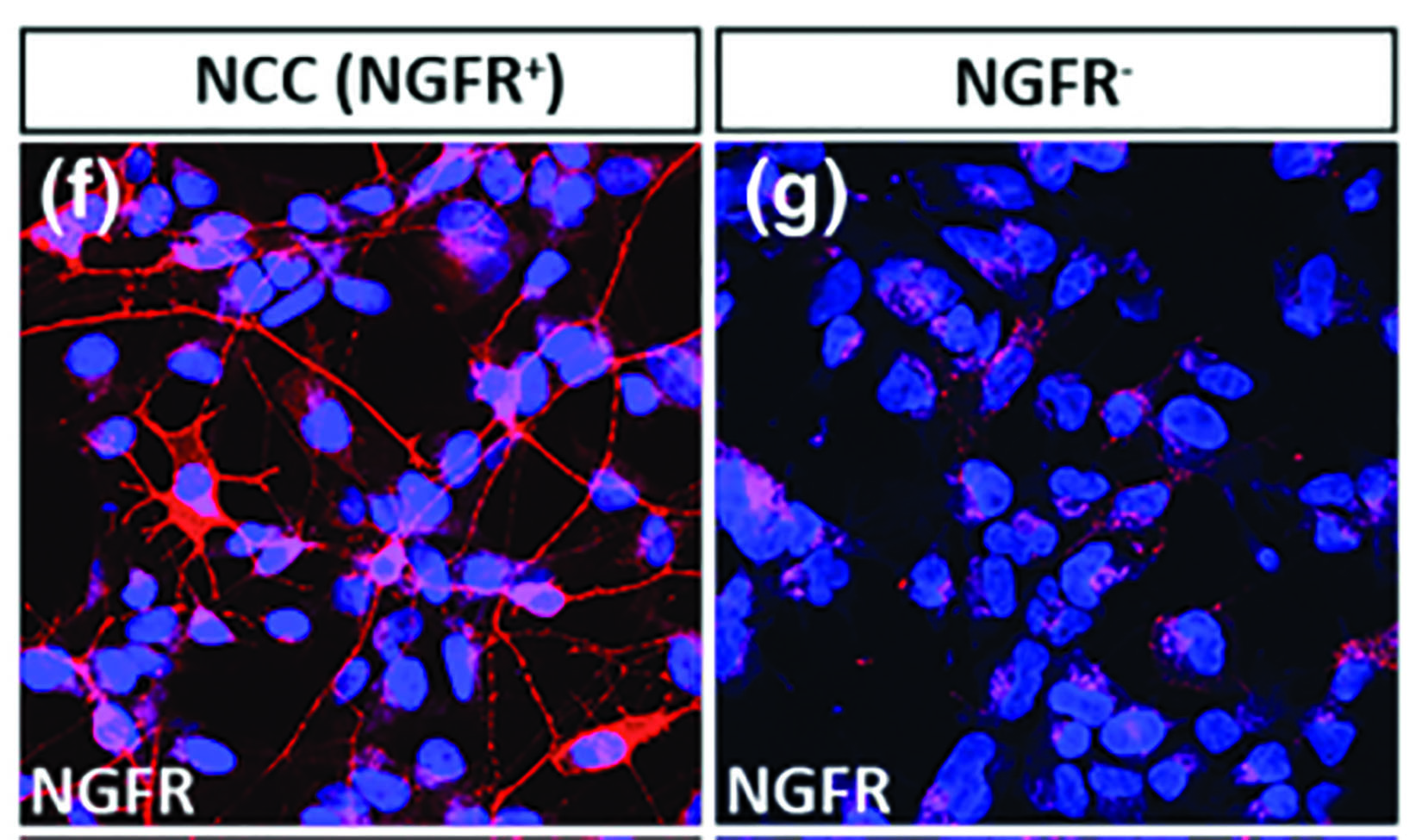
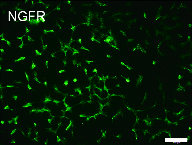
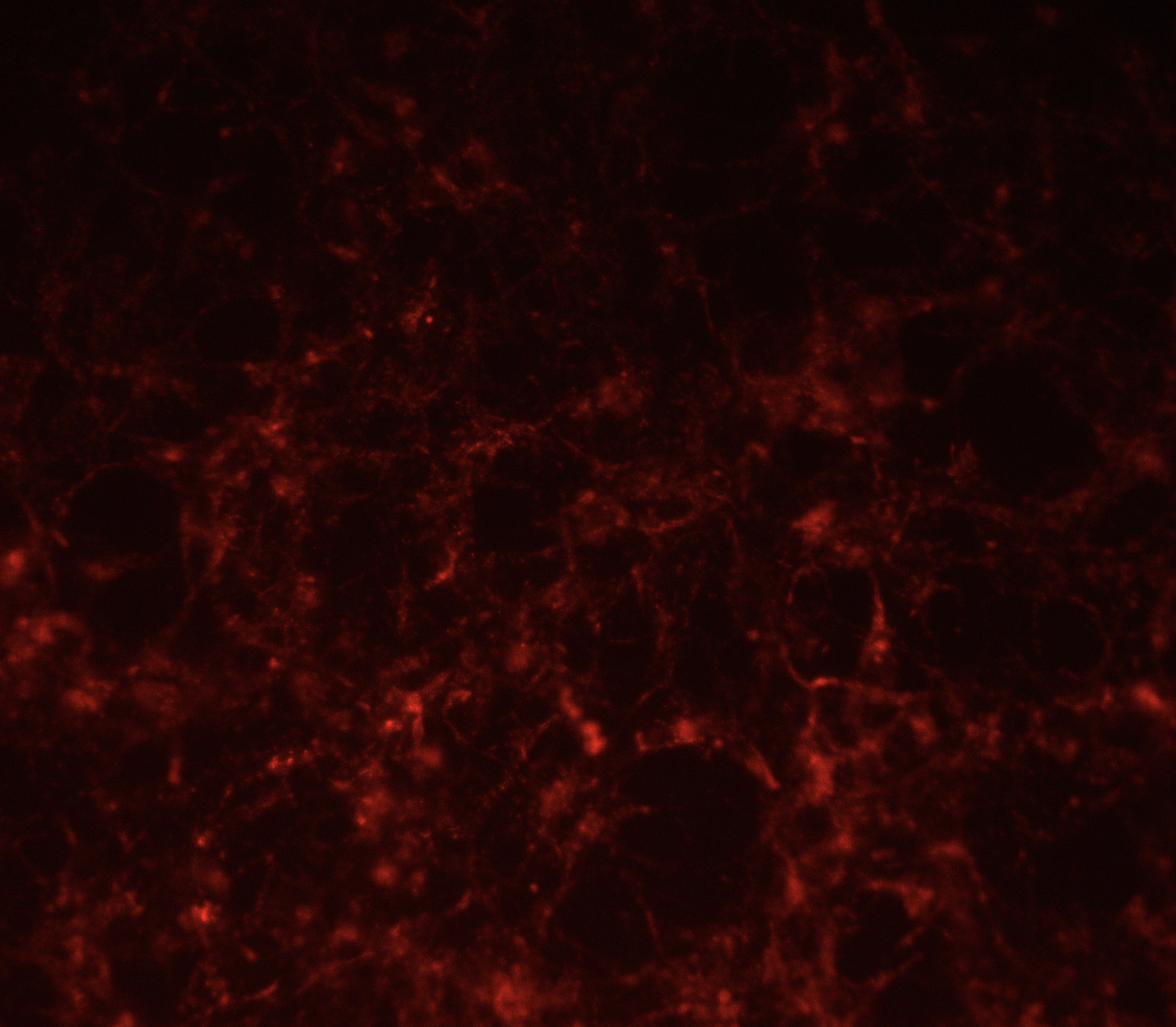
NGFR Antibodies
NGFR (mu p75) Rabbit Polyclonal [AB-N01]
AB-N01 also known as mu p75, and the affinity-purified version, AB-N01AP recognize the p75 receptor in mouse.
Applications include immunohistochemistry, immunoprecipitation, immunoblotting, flow cytometry, and blocking the function of nerve growth factor receptor.
NGFR (ME20.4, p75) Mouse Monoclonal [AB-N07]
AB-N07 also known as ME20.4, recognizes the p75 receptor in human, cat, dog, pig, primate, and sheep.
Applications include flow cytometry, immunoprecipitation, immunohistochemistry, electron microscopy, immunocytochemistry, radioimmunoassay, and targeting.
NGFR (192-IgG, p75) Mouse Monoclonal [AB-N43]
AB-N43 also known as 192-IgG, recognizes the p75 receptor in rat.
Applications include flow cytometry, immunohistochemistry, and targeting.
Melanopsin Antibodies
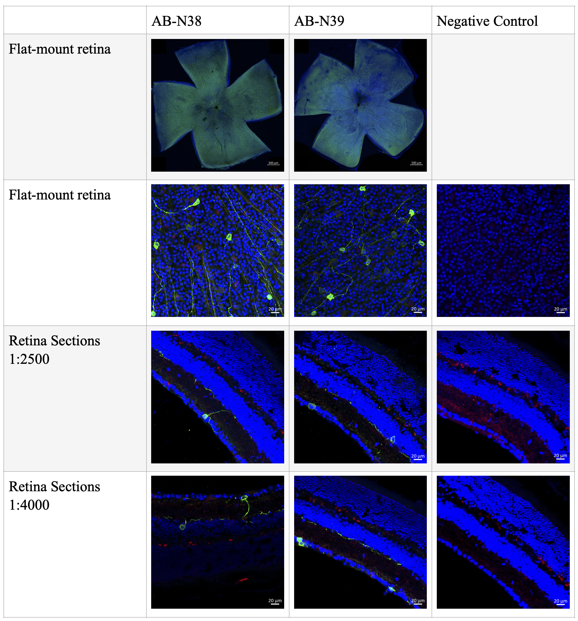
Melanopsin Rabbit Polyclonal [AB-N38]
Anti-Melanopsin (UF006) recognizes a sequence representing the 15 most N-terminal amino acids of the mouse melanopsin extracellular domain. Anti-Melanopsin (UF006) does not cross-react with melanopsins of other species, it is very specific for mouse.
Applications include immunofluorescence.
Melanopsin Rabbit Polyclonal, affinity-purified [AB-N39]
Affinity-purified anti-Melanopsin (UF008) was raised against the 15 N-terminal extracellular amino acids of mouse melanopsin. This antibody does not cross-react with melanopsins of other species, it is very specific for mouse.
Applications include immunofluorescence and targeting. This antibody works in both cross-section and flat-mount staining.