Nakano T, Okita K, Okazaki S, Yoshimoto S, Masuko S, Yagi H, Kato K, Tomioka Y, Imai K, Hamada Y, Masuko K, Shimada-Takaura K, Nagai N, Saya H, Arai T, Ishiwata T, Masuko T (2025) CD44v, S1PR1, HER3, MET and cancer-associated amino acid transporters are promising targets for the pancreatic cancers characterized using mAb. FEBS Open Bio 15(5):867-884. doi: 10.1002/2211-5463.13963 PMID: 39757718
Objective: To analyze pancreatic ductal adenocarcinomas (PDAC) using novel rat mAbs against membrane proteins in conjunction with flow cytometry and immunohistochemistry
Summary: Internalization of membrane proteins by mAbs and growth inhibition by toxin-linked mAbs were demonstrated in many PDAC cell lines, and mAbs against S1PR1, ASCT2, HER3 and CD44v inhibited the growth of xenografted MIA PaCa-2PDAC cells. Furthermore, CD44v-high PDAC showed high mRNA expression of HER1–3, MET and CD44v, and was correlated with poor prognosis. Taken together, the results suggest that CD44v, S1PR1, HER3, MET and the above-mentioned cancer-associated amino acid transporters might be promising targets for the diagnosis and treatment of PDAC.
Usage: Growth inhibition by rat mAbs against PDAC cell lines with secondary antibodies conjugated to saporin (PR-01). Rat mAb solution (4 ug/mL), a PDAC cell suspension and Saporin-conjugated goat anti-rat IgG pAb (Rat-ZAP, IT-26 at 4 ug/mL) were added to each well of 96-well plates.
Park HB, Kim KH, Kim JH, Kim SI, Oh YM, Kang M, Lee S, Hwang S, Lee H, Lee T, Park S, Lee JE, Jeong GR, Lee DH, Youn H, Choi EY, Son WC, Chung SJ, Chung J, Choi K (2024) Improved safety of chimeric antigen receptor T cells indirectly targeting antigens via switchable adapters. Nat Commun 15(1):9917. doi: 10.1038/s41467-024-53996-7 PMID: 39557825
Objective: To show that switchable CAR-T cells with a tumor targeting adaptor can mitigate on-target off-tumor toxicity against a low selectivity tumor antigen that cannot be targeted by conventional CAR-T cells, such as CD40.
Summary: The system is composed of anti-cotinine murine CAR-T cells and cotinine-labeled anti-CD40 single chain variable fragments (scFv), with which the authors show selective tumor killing while sparing CD40-expressing normal cells including macrophages in a mouse model of lymphoma. The authors evaluated whether Cot CAR-T cells could be depleted by Cot-saporin in vivo in an allogeneic CAR-T cell transfer model. When Balb/C mice transplanted with B6 bone marrow cells were injected with B6 Cot CAR-T cells, the transferred Cot CAR-T cells expanded in the peripheral blood in response to Balb/C alloantigen. However, when Cot-saporin was administered during this expansion phase, the Cot CAR-T cells failed to expand and were subsequently eliminated in the blood. Thus, Cot-saporin-mediated CotCAR-T cell suicide was confirmed in vitro and in vivo.
Usage: in vitro Cot CAR-T cell depletion by cotinine-drug conjugates: Peptides were incubated with saporin-labeled streptavidin (IT-27) at a molar ratio of 4:1 to generate cotinine-saporin conjugate (Cot-saporin). For Cot-saporin-dependent cytotoxicity assays on Cot CAR-T cells, a 1:1 mixed population (50,000 cells each) of Cot CAR-T cells (target cells) and control T cells (bystander non-CAR-T cells) were incubated with various doses of Cot-saporin for 48 h in medium containing human IL-2. Seven days after CAR-T cell transfer, Cot-saporin was administered intraperitoneally three times at 3-day intervals.
Jiang H, Cui H, Chen M, Li F, Shen X, Guo CJ, Hoekel GE, Zhu Y, Han L, Wu K, Holtzman MJ, Liu Q (2024) Divergent sensory pathways of sneezing and coughing. Cell 187(21):5981-5997. doi: 10.1016/j.cell.2024.08.009 PMID: 39243765
Objective: To study the difference in sensory receptors and neurotransmission/modulation mechanisms between sneezing and coughing.
Summary: Sneezing and coughing are frequently associated with allergies and respiratory viral infections and it’s assumed both involve common sensory receptors and neurotransmission mechanisms. The author’s work show that the nasal mucosa is innervated by several discrete populations of sensory neurons, but only one population (MrgprC11+MrgprA3−) mediates sneezing. Although this same population innervates the trachea, it does not mediate coughing, and instead, a distinct sensory population (somatostatin SST) mediates coughing but not sneezing. NMB-SAP was used to ablate neruomedin B (NMB) receptor expressing and nucleus tractus solitarius (NTS) neurons. Deletion of these neurons did not affect the coughing responses to Ly344864 and IL-31 (agonists to SST neurons) suggesting that NMB-sensitive NTS neurons do not mediate coughing.
Usage: Neuronal ablation by SST-saporin and NMB-saporin. SST-saporin was made by mixing biotin-labeled somatostatin and Streptavidin-ZAP (IT-27) at a 1:1 molar ratio at room temperature for 20 minutes. SST-Saporin (10 μM, 50 nL), NMB-saporin (#IT-70; 50 ng in 50 nL) or Blank-SAP (#IT-21; 10 μM in 50 nL or 50 ng in 50 nL) was injected into the NTS region.
O’Sullivan JA, Youngblood BA, Schleimer RP, Bochner BS (2024) Siglecs as potential targets of therapy in human mast cell- and/or eosinophil-associated diseases. Semin Immunol 69:101799. doi: 10.1016/j.smim.2023.101799 PMID: 37413923
Objective: To review a subset of Siglecs and their various endogenous or synthetic sialoside ligands that regulate eosinophil and mast cell function and survival.
Summary: Sialic acid-binding immunoglobulin-like lectins (Siglecs) are vertebrate glycan-binding cell-surface proteins. Many Siglecs mediate cellular inhibitory activity and are of interest as part of a strategy to therapeutically lessen unwanted cellular responses. Human eosinophils and mast cells express overlapping but distinct patterns of Siglecs, and certain Siglecs have become the focus of novel therapies for allergic and other eosinophil and mast cell-related diseases.
Usage: Saporin in conjunction with CD22 glycomimetic ligand BPCNeuAc leads to cells death induction in a ligand-dependent manner on B-lymphoma cells (Collins et al.). Incubation with anti-Siglec-8 monoclonal antibody conjugated to saporin led to the death of malignant mast cells and eosinophils (O’Sullivan et al.)
Bryder D, Konturk-Ciesla A, Zhang Q, Kharazi S (2024) A non-invasive stem cell therapy boosts lymphopoiesis and averts age-related blood diseases in mice. Research Square doi: 10.21203/rs.3.rs-4528815/v1
Objective: To develop a murine transplantation model based on low-intensity conditioning protocols using antibody-mediated Hematopoietic stem cell.
Summary: Authors demonstrate that young HSCs, once transplanted, survive and thrive in aged hosts, dramatically improving hematopoietic output and ameliorating age-compromised lymphopoiesis.
Usage: Intravenous injection of CD45-SAP (3 mg/kg, IT-91). Biotinylated anti-CD45.2 antibodies were mixed with streptavidin-saporin conjugate at a 1:1 molar ratio (IT-27).
Garaudé S (2024) Engineering an antibody-discernible and functional CD45 variant on hematopoietic stem and progenitor cells via base editing. Univ Basel Thesis.
Objective: To develop a system for a cell surface marker present on all hematopoietic cell to deplete malignant cells. Authors screened CD45’s extracellular domain regions with base editors and generated multiple CD45 protein variants altering the binding of antibodies.
Summary: Authors selected the CD45 K352E/G variant profile and improved its base editing rate as it showcased loss of binding of a unique anti-CD45 antibody while still maintaining the surface marker’s expression, stability, and function in human hematopoietic stem and progenitor cells (HSPCs). The resulting loss of antibody binding prompted the modification and humanization of the anti-CD45 antibody, culminating in the development of an anti-CD45 antibody-drug conjugate (CD45-ADC; CIM053-SG3376). In an AML mouse models xenografted with HSPCs, administration of the CD45-ADC selectively depleted human leukemia and HSPCs wt cells while preserving the healthy hematopoietic system derived from the transplanted base edited HSPCs.
Usage: In vitro antibody-drug-conjugate (ADC) mediated killing assays. For ADC killing assays involving saporin, a 100 nM stock was prepared by incubating the biotinylated antibody and Streptavidin-ZAP at a 1:1 molar ratio for 30 min at room temperature
Garaudé S, Marone R, Lepore R, Devaux A, Beerlage A, Seyres D, Dell’ Aglio A, Juskevicius D, Zuin J, Burgold T, Wang S, Katta V, Manquen G, Li Y, Larrue C, Camus A, Durzynska I, Wellinger LC, Kirby I, Van Berkel PH, Kunz C, Tamburini J, Bertoni F, Widmer CC, Tsai SQ, Simonetta F, Urlinger S, Jeker LT (2024) Selective haematological cancer eradication with preserved haematopoiesis. Nature 630(8017):728-735. doi: 10.1038/s41586-024-07456-3 PMID: 38778101
Objective: To demonstrate that an antibody–drug conjugate (ADC) targeting the pan-haematopoietic marker CD45 enables the antigen-specifc depletion of the entire haematopoietic system, including Haematopoietic stem cells ( HSC).
Summary: Pairing this ADC with the transplantation of human HSCs engineered to be shielded from the CD45-targeting ADC enables the selective eradication of leukaemic cells with preserved haematopoiesis. The combination of CD45-targeting ADCs and engineered HSCs creates an almost universal strategy to replace a diseased haematopoietic system, irrespective of disease aetiology or originating cell type.
Usage: For ADC killing assays involving saporin, a 100 nM stock was prepared by incubating the biotinylated antibody (BC8 or MIRG451 mAbs) and saporin–streptavidin (IT-27) at a 1:1 molar ratio for 30 min at room temperature
Simpson J, Starke CE, Ortiz AM, Ransier A, Darko S, Llewellyn-Lacey S, Fennessey CM, Keele BF, Douek DC, Price DA, Brenchley JM (2024) Immunotoxin-mediated depletion of Gag-specific CD8+ T cells undermines natural control of Simian immunodeficiency virus. JCI Insight e174168. doi: 10.1172/jci.insight.174168 PMID: 38885329
Objective: To investigate the role of gag epitope-specific CD8+ T cells in the immune control of Simian Immunodeficiency Virus (SIV) in a nonhuman primate model.
Summary: Antibody-mediated depletion studies suggest that CD8+ T cells suppress SIV replication, but bulk depletion of CD8+ T cells may increase SIV target cells. Authors selectively depleted CD8+ T cells specific to the CM9 epitope, but this didn’t suppress viral replication in SIV-infected rhesus macaques. The data indicate that CM9-specific CD8+ T cells alone are not sufficient for immune control of SIV.
Usage: Streptavidin-ZAP was added stepwise to purified CM9 monomers to a final molar ratio of 1:4 and administered intravenously at a doses of 350 pmol/kg, 500 pmol/kg, 1 nmol/kg, or 2 nmol/kg at various time intervals.
Hello, I am very interested in using your Fab-ZAP line. So Fab-ZAPs simply tells us what mAbs internalize best – is that right? Can you send me some example data? I’d love to see a cell line that has been evaluated and the data that has been generated.
Answer:
Yes, Fab-ZAPs will tell you what mAbs internalize best. We have a variety of different versions of these products (ZAP secondary conjugates) which you can find here.
These have been tested on many different cell lines, both in-house and by our customers.
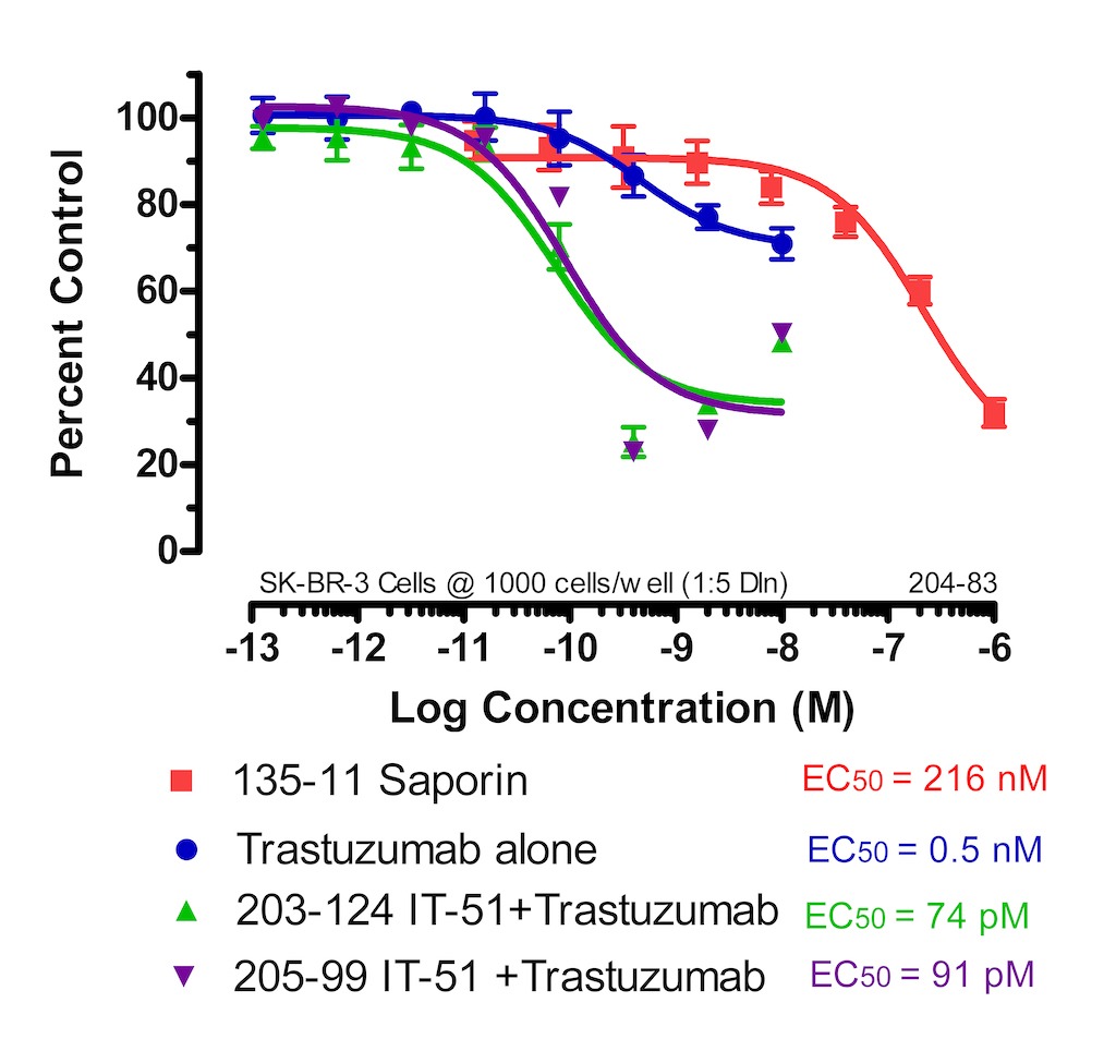
I’ve included a graph that corresponds to Fab-ZAP human (IT-51). Here is a cytotox which tests Fab-ZAP human with Trastuzumab on SK-BR-3 cells (breast cancer cell line).
SK-BR-3 cells were plated at 1000 cells/90 μl/well and incubated overnight. Trastuzumab and Saporin dilutions were made in cell media and 10 μl was added to each well. The Trastuzumab was also diluted in cell media containing, at a final concentration, 4.5 nM/10 μl Fab-ZAP, and 10 μl was added to each well. The plates were incubated for 72 hours. The plates were developed using a solution of XTT/PMS and read at 450 nm. Cytotoxicity was analyzed by comparing well readings of the treated wells to those of the control wells, expressed as a percentage. The number of viable cells remaining on the day of development is measured via cell metabolism of a colorimetric molecule within the developing reagents. Analysis was performed using Prism software (GraphPad, San Diego).
Related Products
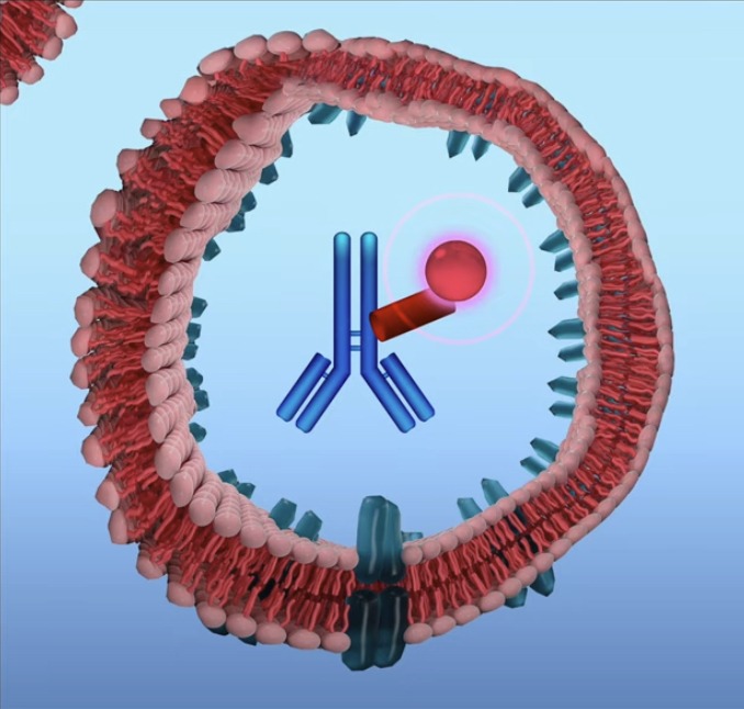
ZAP conjugates – These are non-targeted saporin conjugates that “piggyback” on to your primary targeting agent (biotinylated material or antibody) to eliminate specific cells and reveal cell function.
Where does the saporin payload release after internalization? For example, does it require trafficking into a late endosome/lysosomal compartment?
Answer:
Thank you for reaching out to us. Hopefully I can help answer some of your questions regarding what happens to saporin after being internalized.
I would first like to refer you to an article we published, titled “Streptavidin-Saporin: Converting Biotinylated Materials into Targeted Toxins”. In it we review the internalization of saporin and include a few references for support. To answer your question in general, yes the conjugate is typically endocytosed and makes its way to the late endosome.
As an overview of this debated topic, the Wensley, H.J. et al 2019 article (ref #4) studied the escape of saporin from the late endosome and examined the endocytic process to quantify the endosomal escape into the cytosol. The Holmes, S.E et al 2015 (ref #5) and Giansanti, F. et al 2018 (ref #6) articles describe additional research examining chemical and genetic strategies used in assisting in saporin’s escape from the endosome. After endocytocis, Vago, R. et al 2005 (ref #7) compared saporin and ricin A chain and other bacterial toxins looking at their different intracellular routes to enter the cytosol. These articles should provide a nice foundation and hopefully better answer any questions.
If you’re interested in visualizing lysosomal trafficking, you might consider our pHast product line. These are secondary pH-dependent fluorescent conjugates, meaning that they only fluoresce once inside the endosomes and lysosomes of cells (which are acidic compared to the cytosol).
Related Products
pHast Conjugates – one of our pHastest tools for quantitative testing.