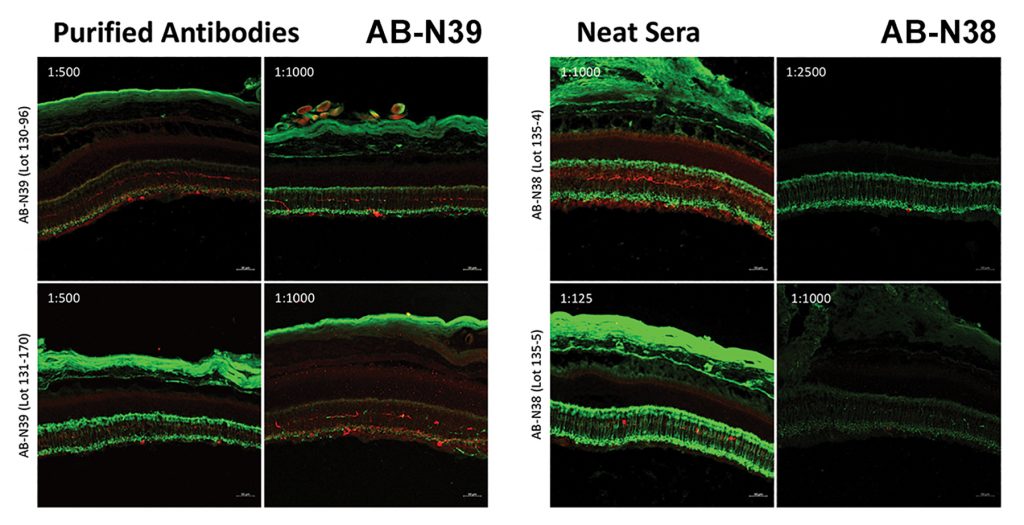
In 1997, Melanopsin was discovered by Dr. Ignacio Provencio as a novel opsin in the melanophores (light-sensitive skin cells) of the African clawed frog.1 Provencio and colleagues then found in 2000 that melanopsin is also present in mouse retina, specifically in ganglion cells, and that it mediates non-visual photoreceptive tasks.2 The figure below illustrates the specificity of both the Melanopsin Rabbit Antisera [AB-N38] and the affinity purified version [AB-N39].
- Provencio I, Jiang G, De Grip WJ, Hayes WP, & Rollag MD. Melanopsin: An opsin in melanophores, brain, and eye. (1998). Proceedings of the National Academy of Sciences, 95 (1):340.
- Provencio I, Rodriguez IR, Jiang G, Hayes WP, Moreira EF, & Rollag MD. A Novel Human Opsin in the Inner Retina. (2000). The Journal of Neuroscience, 20 (2):600.
Recent References using Anti-Melanopsin
Quattrochi LE, Stabio ME, Kim I, Ilardi MC, Michelle Fogerson P, Leyrer ML, & Berson DM. The M6 Cell: A Small-Field Bistratified Photosensitive Retinal Ganglion Cell. (2019). J Comp Neurol, 527 (1):297-311.
[AB-N38] Summary: M6 cells express low levels of melanopsin and have correspondingly weak intrinsic light responses.
Dose: In all experiments involving melanopsin immunofluorescence, melanopsin immunodetection was enhanced using tyramide signal amplification coupled with horseradish peroxidase (HRP)-paired with goat anti-rabbit IgG and an Alexa fluorophore. 1:10,000.
Sweeney NT, James KN, Nistorica A, Lorig-Roach RM, & Feldheim DA. Expression of Transcription Factors Divides Retinal Ganglion Cells into Distinct Classes. (2019). J Comp Neurol, 527 (1):225-235.
[AB-N38] Dose: Melanopsin (Opn4) Immunostaining 1:1000.