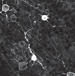
Synergistic loss of interneurons and RGCs in Pcdh-ag double mutant retinas. “…surviving RGC subpopulations are sparse, but proportionally more MelipRGCs remained in mutant Pcdh-agrko retinas (4.1% of total RGCs are Melanopsin+ in Control compared with 35% of Melanopsin+ /total RGCs in Pcdh-agrko mutants).”
In 1997, Melanopsin was discovered by Dr. Ignacio Provencio as a novel opsin in the melanophores (light-sensitive skin cells) of the African clawed frog.[1] Provencio and colleagues then found in 2000 that melanopsin is also present in mouse retina, specifically in ganglion cells, and that it mediates non-visual photoreceptive tasks.[2] Melanopsin has since been found to be encoded by Opn4 with orthologs in a variety of organisms. Here are publications from 2018 using Anti-Melanopsin (Cat. #AB-N38, Cat. # AB-N39) and Melanopsin-SAP (Cat. #IT-44) to further explore the roles of melanopsin in various species.
- Provencio I, Jiang G, De Grip WJ, Hayes WP, & Rollag MD. Melanopsin: An opsin in melanophores, brain, and eye. (1998). Proceedings of the National Academy of Sciences, 95 (1):340.
- Provencio I, Rodriguez IR, Jiang G, Hayes WP, Moreira EF, & Rollag MD. A Novel Human Opsin in the Inner Retina. (2000). The Journal of Neuroscience, 20 (2):600.
- Ing-Esteves S, Kostadinov D, Marocha J, Sing AD, Joseph KS, Laboulaye MA, Sanes JR, & Lefebvre JL. Combinatorial Effects of Alpha- and Gamma-Protocadherins on Neuronal Survival and Dendritic Self-Avoidance. (2018). The Journal of Neuroscience, 38 (11):2713. Cat. #AB-N38: Anti-Melanopsin
- Delwig A, Chaney SY, Bertke AS, Verweij JAN, Quirce S, Larsen DD, Yang C, Buhr E, Van Gelder R, Gallar J, Margolis T, & Copenhagen DR. Melanopsin expression in the cornea. (2018). Visual Neuroscience, 35 E004. 01/31. Cat. #AB-N39: Anti-Melanopsin
- Grillo SL, & Stella SL, Jr. Melanopsin retinal ganglion cells are not labeled in Thy-1YFP-16 transgenic mice. (2018). Neuroreport, 29 (2):118-122. 2017/12/19. Cat. #AB-N39: Anti-Melanopsin
- Potter H, Alenciks E, Frazier K, Porter A, & Fraley GS. Immunolesion of melanopsin neurons causes gonadal regression in Pekin drakes (Anas platyrhynchos domesticus). (2018). Gen Comparative Endocrinology, 256 16-22. Cat. #IT-44; Melanopsin-SAP, Cat. # PR-01; Saporin
- Stabio ME, Sabbah S, Quattrochi LE, Ilardi MC, Fogerson PM, Leyrer ML, Kim MT, Kim I, Schiel M, Renna JM, Briggman KL, & Berson DM. The M5 Cell: A Color-Opponent Intrinsically Photosensitive Retinal Ganglion Cell. (2018). Neuron, 97 (1):150-163.e154. Cat. #AB-N38: Anti-Melanopsin
- Ueki Y, Shchepetkina V, & Lefcort F. Retina-specific loss of lkbkap/Elp1 causes mitochondrial dysfunction that leads to selective retinal ganglion cell degeneration in a mouse model of familial dysautonomia. (2018). Dis Model Mech. Jul 30;11(7). pii: dmm033746. doi: 10.1242/dmm.033746. Cat. #AB-N38: Anti-Melanopsin
- Wong JCY, Smyllie NJ, Banks GT, Pothecary CA, Barnard AR, Maywood ES, Jagannath A, Hughes S, van der Horst GTJ, MacLaren RE, Hankins MW, Hastings MH, Nolan PM, Foster RG, & Peirson SN. Differential roles for cryptochromes in the mammalian retinal clock. (2018). FASEB J Aug;32(8):4302-4314. doi: 10.1096/fj.201701165RR. Epub 2018 Mar 21. Cat. #AB-N38; Anti-Melanopsin