3D Visualization of Individual Regenerating Retinal Ganglion Cell Axons Reveals Surprisingly Complex Growth Paths.
Bray, ER, Noga, M, Thakor, K, et al. (2017). eNeuro eNeuro doi: 10.1523/ENEURO.0093-17.2017 PMID: 28856242
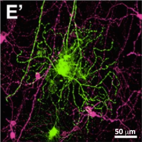
“Our study demonstrates extensive and circuitous RGC axon elongation both in pre- and post-lesion regions, highlighting the need to better understand the factors that inhibit direct axon growth in the optic nerve.”
Immunohistochemistry: Rabbit-anti-melanopsin (OPN4; UF006) 1:2500 (Cat. #AB-N38)
Also see:
Somasundaram P, Wyrick GR, Fernandez DC, Ghahari A, Pinhal CM, Simmonds Richardson M, Rupp AC, Cui L, Wu Z, Brown RL, Badea TC, Hattar S, Robinson PR. (2017) C-Terminal Phosphorylation Regulates the Kinetics of a Subset of Melanopsin-Mediated Behaviors in Mice. Proc Natl Acad Sci U S A 114(10):2741-46. PMID: 28223508 (Targeting Trends)
Gonzalez-Menendez I, Contreras F, Cernuda-Cernuda R, Provencio I, Garcia-Fernandez JM (2010) Postnatal development and functional adaptations of the melanopsin photoreceptive system in the albino mouse retina. Invest Ophthalmol Vis Sci 51(9):4840-4847. (Targeting Trends)
Göz D, Studholme K, Lappi DA, Rollag MD, Provencio I, Morin LP (2008) Targeted destruction of photosensitive retinal ganglion cells with a saporin conjugate alters the effects of light on mouse circadian rhythms. PLoS ONE 3(9):e3153. (Targeting Trends)
Provencio I, Rollag MD, Castrucci AM (2002) Photoreceptive net in the mammalian retina. Nature 415:493.
(Anti-Melanopsin; Cat. #AB-N38)
Opsin-targeted research tools
Melanopsin-SAP [IT-44]
Melanopsin Rabbit Polyclonal [AB-N38]
Melanopsin Rabbit Polyclonal, affinity-purified [AB-N39]
RGR-opsin Rabbit Polyclonal [AB-N45]