Leduc-Pessah H, Weilinger N, Fan C, Burma N, Thompson R, Trang T (2017) Site-specific regulation of P2X7 receptor function in microglia gates morphine analgesic tolerance. J Neurosci 37:10154-10172.. doi: 10.1523/JNEUROSCI.0852-17.2017
Summary: By selectively ablating microglia in the spinal cord using a Mac-1-SAP the authors demonstrate a causal role for microglia in the development, but not maintenance, of morphine tolerance in male rats.
Usage: Mac-1-SAP or unconjugated Saporin control (15 μg) was administered by intrathecal injection.
Capone E, Giansanti F, Ponziani S, Lamolinara A, Iezzi M, Cimini A, Angelucci F, Sorda R, Laurenzi V, Natali PG, Ippoliti R, Iacobelli S, Sala G (2017) EV20-Sap, a novel anti-HER-3 antibody-drug conjugate, displays promising antitumor activity in melanoma. Oncotarget 8(56):95412-95424. doi: 10.18632/oncotarget.20728 PMID: 29221137
Objective: Using EV20 (Anti-Her3)-SAP to target Her3 positive melanoma
Summary: Linking a humanized monoclonal antibody targeting Her3, named EV20, to saporin creates a powerful cytotoxic agent targeting melanoma – which overexpress the Her3 receptor. The antibody, EV20, rapidly is internalized by Her3 whether its dependent ligand is there or not. EV20-SAP demonstrated potent and specific cytotoxicity towards human melanoma cells.
Usage: At a concentration of 19 nM only 10% of the human melanoma cells 9 (SK-MEL24 melanoma cells) survived, where free saporin at the same concentration shows no/minimal cytotoxicity.
Echeverry S, Shi XQ, Yang M, Huang H, Wu Y, Lorenzo L-E, Perez-Sanchez J, Bonin RP, De Koninck Y, Zhang J (2017) Spinal microglia are required for long-term maintenance of neuropathic pain. Pain 158:1792-1801.. doi: 10.1097/j.pain.0000000000000982
Summary: Selective depletion of spinal microglia in male rats with the targeted immunotoxin Mac-1-SAP and blockade of brain derived neurotrophic factor–TrkB signalling with intrathecal TrkB Fc chimera, but not cytokine inhibition, almost completely reversed pain hypersensitivity. To selectively deplete microglia in the spinal cord, Mac-1-SAP was injected intrathecally. In each group, rats received either an intrathecal injection of 12 mg/7 mL of Mac-1-SAP (n = 6-8) or equal volume of 0.9% saline (n 5 6) or the inactive unconjugated toxin, Saporin (n = 6).)
Burma NE, Bonin RP, Leduc-Pessah H, Baimel C, Cairncross ZF, Mousseau M, Shankara JV, Stemkowski PL, Baimoukhametova D, Bains JS, Antle MC, Zamponi GW, Cahill CM, Borgland SL, De Koninck Y, Trang T (2017) Blocking microglial pannexin-1 channels alleviates morphine withdrawal in rodents. Nat Med 23:355-360.. doi: 10.1038/nm.4281
Summary: The authors investigated the mechanisms underlying opiate withdrawal in rat. Depletion of spinal lumbar microglia by intrathecal injections of Mac-1–SAP (Cat. #IT-33; 20 mcg) decreased withdrawal behaviors and attenuated the severity of withdrawal without affecting morphine antinociception. Unconjugated Saporin (Cat. #PR-01; 20 mcg) was used as control and had no effect on spinal CD11b immunoreactivity or naloxone-induced morphine withdrawal.
Polito L, Djemil A, Bortolotti M (2016) Plant toxin-based immunotoxins for cancer therapy: a short overview. Biomedicines 4(2):12. doi: 10.3390/biomedicines4020012
Lee S, Diener K, Kaufman S, Krieger J, Pettersen K, Jejelava N, Arnold M, Watts A, Langhans W (2016) Limiting glucocorticoid secretion increases the anorexigenic property of Exendin-4. Mol Metab 5:552-565. doi: 10.1016/j.molmet.2016.04.008
Summary: Glucagon-like peptide-1 (GLP-1) analogs lower blood surgar levels and cause a loss of appetite. Exendin-4 (Ex-4) is a GLP-1 receptor agonist, and also increases glucocorticoid secretion. Several tests were conducted to determine if the released glucocorticoids interact with Ex-4’s anorexigneic effect. One method involved ablating hindbrain catecholaminergic neurons by stereotaxically injecting 42 ng of Anti-DBH-SAP (Cat. #IT-03) bilaterally into the paraventricular nucleus of the hypothalamus in rats. Animals were injected with equimolar concentrations of unconjugated Saporin (Cat. #PR-01) as a control. Anti-DBH-SAP lesions reduced the efficacy of Ex-4 to increase corticosterone secretion but increased the anorexigenic effect, indicating that Ex-4-dependent corticosterone secretion opposes Ex-4’s actions. Anti-DBH-SAP lesions increased Ex-4’s ability to reduce food intake and body weight.
Sedlik C, Heitzmann A, Viel S, Ait Sarkouh R, Batisse C, Schmidt F, De La Rochere P, Amzallag N, Osinaga E, Oppezzo P, Pritsch O, Sastre-Garau X, Hubert P, Amigorena S, Piaggio E (2016) Effective antitumor therapy based on a novel antibody-drug conjugate targeting the Tn carbohydrate antigen. Oncoimmunology 5:e1171434. doi: 10.1080/2162402X.2016.1171434
Summary: Scientists wanted to study the potential of Chi-Tn, a monoclonal antibody against a glycol-peptidic tumor-associated antigen, as an anticancer antibody-drug conjugate. They demonstrated that Chi-Tn specifically targeted tumor cells in vivo, using flow cytometry and deconvolution microscopy to show that Chi-Tn is rapidly internalized. Chi-Tn-SAP (ATS Custom Services) effectively killed Tn-positive cells, but had no effect on Tn-negative cells. Saporin (Cat. #PR-01) was used as control. The cytotoxicity of the Chi-Tn-SAP correlated with the level of tumoral Tn expression.
Over the years, ATS has frequently been asked about Saporin’s safety for use in the lab as well as when used clinically. Residual awareness of alternate Ribosome-Inactivating Proteins (RIPs) and ‘toxins’ such as Ricin have caused some researchers new to the use of RIPs to question the belief that Saporin is safe. Unlike Type 2 RIPs (such as Ricin), Type I RIPs, like Saporin have no binding chain and consequently no means of entering the physiological space necessary for the protein to act as a toxin. The following is a review of safety in handling and potential toxicity within the human body for systemic events not related to normal research applications of Saporin conjugates, including Substance P-Saporin (SP-SAP), which is a therapeutic under development for the treatment of chronic pain.
The acute LD50 for saporin in mice (25 g) is 6.8 mg/kg;[1] that would translate in humans (75 kg) to 510 mg! A concentration of about 100 nM is the threshold to see even a vague hint of saporin toxicity. In human blood, that would correspond to 24 mg injected systemically into a person. The fermentation process to produce recombinant saporin has a titer of 2 mg/L meaning that the production broth itself contains no more than 67 nM concentration of saporin. Furthermore, the final protein concentrations from production batches of recombinant Saporin used in our drug are 4 mg/ml, meaning 6 mL of final material would need to accidentally end up in a human before the ‘hint of toxicity’ threshold would potentially be met.
The toxicology studies of SP-SAP contained within ATS’s IND prior to the current human Phase I clinical trial evaluated effects related to the intended method of administration, intrathecal local injection. SP-SAP is not expected to ever be a self-administered therapy, so the effects of gross off-target events, such as accidental auto-injection, swallowing, spillage, or immersion were not considered.
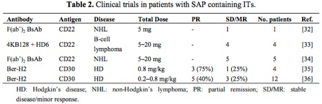
The table below[2] highlights antibody-saporin conjugates approved by the FDA for Phase I/II clinical trials in humans. The therapeutics listed below were administered intravenously and imply what the FDA accepted as non-toxic levels of saporin-based conjugates in these studies.
Looking more closely at the study by French et al.,[3] several milligrams of antibody conjugate were repeatedly injected into human patients under a FDA regulated clinical trial and peak serum levels tested, demonstrating rapid clearing of saporin from the system.
As a company that specializes in Saporin, our two-plus decades of experience working with the protein in research, preclinical, and clinical environments has taught us that with minimal standard laboratory precautions users are not at any real risk of toxic effects. Even our CSO, after 30+ years of working with Saporin exhibits undetectable levels of Saporin antibodies in his blood!
Gritsch S, Bali K, Kuner R, Vardeh D (2016) Functional characterization of a mouse model for central post-stroke pain. Mol Pain 12:1744806916629049. doi: 10.1177/1744806916629049
Summary: While clinical evidence has pointed toward central pain pathway dysfunction in central post-stroke pain (CPSP), the underlying mechanisms have not been defined. In this work the authors created a mouse model of CPSP through lesions of the thalamic ventral posterolateral nucleus. In order to examine the role of neurokinin-1 receptor-expressing (NK1R) neurons in lamina I/III of the spinal cord in the development and maintenance of CPSP the authors administered 1 μmol intrathecal injections of SSP-SAP (Cat. #IT-11). Saporin (Cat. #PR-01) was used as a control. While the NK1R+ neurons in the spinal cord were not involved in establishing CPSP, the data indicate that sensory changes in the mice are comparable to those observed in human patients with CPSP.