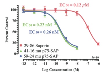
The cover article this quarter demonstrates usage of mu p75-SAP (Cat. #IT-16) to study delirium. To create this toxin, we affinity-purified the rabbit polyclonal to the low affinity neurotrophin receptor, p75, (Cat. #AB-N01AP) with the immunogen bound to a solid support, and then conjugated the affinity-purified antibody to saporin (Cat. #PR-01). As can be seen in the cytotoxicity assay below, mu p75-SAP has an EC50 in the nanomolar range. This high potency translates to smaller amounts used for elimination of p75-positive neurons in the mouse brain which results in a greater index of efficacy and lesser non-specific cytotoxicity.
The mu p75-SAP kit includes, in addition to the immunotoxin, equal aliquots of saporin (Cat. #PR-01), the affinity-purified rabbit polyclonal antibody (Cat. #AB-N01AP), and the control immunotoxin, Rabbit-IgG-SAP (Cat. #IT-35).
Also available are fluorescent conjugates of AB-N01AP: Cy3-labeled Anti-murine NGFr (Cat. #AB-N01APFL3), and Cy5-labeled Anti-murine NGFr (Cat. #AB-N01APFL5).
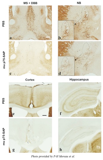
(Fig 1 from Moreau et al. Targeting Trends, 9(2): pp. 1,6, 2008.)