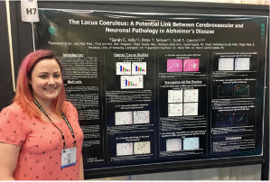
This year’s Society for Neuroscience meeting in San Diego offered over 30,000 people the chance to discuss the latest discoveries and trends in the field. ATS products were well represented in 20 posters on a variety of topics. Though there were many excellent candidates, the committee felt the work done by Kelly et al from Michigan State Univ. exemplified the innovative approaches and conclusions that can be reached using targeted toxins, such as Anti-DBH-SAP (CAT# IT-03), and was awarded our Poster of the Year.
Be sure to check out the featured article in Targeting Trends.
Noradrenergic locus coeruleus (LC) neuron loss is a major feature of Alzheimer’s disease (AD). The LC is the primary source of norepinephrine (NE) in the forebrain, where it modulates attention and memory in vulnerable cognitive regions such as prefrontal cortex and hippocampus. Furthermore, LC-mediated NE signaling is thought to play a role in blood brain barrier maintenance and neurovascular coupling, suggesting that LC degeneration may impact the high comorbidity of cerebrovascular disease (CVD) and AD. However, the extent to which LC projection system degeneration occurs in the earliest stages of AD is not fully characterized to date. To address these issues, we analyzed LC tissue samples from University of Kentucky AD Center subjects who died with a premortem diagnosis of no cognitive impairment (NCI) and Braak stages 0-II at autopsy, NCI subjects with Braak stages III-V thought to be in a preclinical AD (PCAD) stage, and subjects with mild cognitive impairment (MCI) or mild AD (n = 5-6 cases/group). Paraffin-embedded pontine tissue blocks containing the LC were cut at 20µm, immunostained with tyrosine hydroxylase (TH, a marker for NE synthesis), and analyzed by stereology to estimate total LC neuron number (total number of neuromelanin-containing LC neurons) and the percentage of TH+ LC neurons. Preliminary analysis reveal a ~20% loss of both total and TH+ LC neurons in PCAD (p = 0.08), a ~30-35% loss of these neurons in MCI (p < 0.05), and a ~45-50% loss of total and TH+ neurons in AD (p < 0.01) compared to NCI. Studies were also performed to compare additional LC neuronal pathologies (phospho-tau, TDP-43, and 8dOHG) in the diagnostic groups. A substantial increase in 8dOHG and phospho-tau is observed in PCAD compared to NCI. The morphometric data will be correlated with postmortem neuropathologic and CVD variables (e.g., microinfarcts and cerebral amyloid angiopathy) to gauge the relationship between LC neurodegeneration and cerebral AD and vascular pathology. To model these relationships in vivo, we stereotactically lesioned LC projection neurons innervating the PFC, a major LC projection zone, in the TgF344-19 rat model of AD (6 months old) using the noradrenergic immunotoxin, dopamine-β-hydroxylase-saporin, or a control lesion (n = 8/group). Prior to sacrifice at 9 months, immunotoxin- and control-lesioned rats will be tested behaviorally on the Barnes maze task. Postmortem PFC will be analyzed for LC fiber innervation, NE and NE metabolite levels, CVD pathology and AD-like pathology. Taken together, these data will shed light on the multifactorial noradrenergic pathways contributing to neuronal and vascular pathologies during the onset of AD.