Cholecystokinin (CCK) is widely distributed in the central nervous system and the gastrointestinal tract. The 33-amino acid peptide contains a carboxyl terminal octapeptide sequence Asp-Tyr-Met-Gly-Trp-Met-Asp-Phe-NH2 which confers the biological activity of CCK, and where the tyrosine residue occurs in sulfated form. This octapeptide, CCK8(SO3), has high affinity for the two structurally-defined CCK receptors types, CCK1 and CCK2.
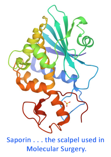
CCK-SAP is a chemical conjugate of sulfonated CCK and the ribosome-inactivating protein, saporin. It specifically eliminates cells that express CCK Receptor.
CCK-SAP is available individually (Cat. #IT-31) or as a kit (Cat. #KIT-31) which includes CCK-SAP and Blank-SAP (Cat. #IT-21).
keywords: cholecystokinin, CCK, CCK1, CCK2, CCK receptor, sulfonated CCK, saporin, central nervous system, gastrointestinal tract, eating, appetite, brain, neuroscience, peptide, octapeptide, CNS, gut, nodose, vagal, obesity, pain, ganglia
Neuropathic pain is maintained by brainstem neurons co-expressing opioid and cholecystokinin receptors.
Zhang W, Gardell S, Zhang D, Xie JY, Agnes RS, Badghisi H, Hruby VJ, Rance N, Ossipov MH, Vanderah TW, Porreca F, Lai J (2009) Neuropathic pain is maintained by brainstem neurons co-expressing opioid and cholecystokinin receptors. Brain 132:778-787. doi: 10.1093/brain/awn330
Summary: It has been hypothesized that a subset of rostral ventromedial medulla (RVM) neurons co-expressing the cholecystokinin type 2 receptor and the mu-opioid receptor are responsible for the maintenance of neuropathic pain. Rats were treated with 50-ng bilateral RVM injections of Dermorphin-SAP (Cat. #IT-12), CCK-SAP (Cat. #IT-31), or saporin (Cat. #PR-01) as a control. Lesion of the RVM neurons prevented hyperalgesia in response to CCK treatment, and shortened abnormal pain states caused by sciatic nerve injury.
Related Products: Dermorphin-SAP / MOR-SAP (Cat. #IT-12), CCK-SAP (Cat. #IT-31), Saporin (Cat. #PR-01)
Lumbar intrathecal CCK-saporin: anatomic and nociceptive effects
Datta S, Chatterjee K, Kline IV RH, Wiley RG (2008) Lumbar intrathecal CCK-saporin: anatomic and nociceptive effects. Neuroscience 2008 Abstracts 773.4/MM32. Society for Neuroscience, Washington, DC.
Summary: Lumbar intrathecal CCK (cholecystokinin) appears anti-opiate in nocifensive reflex testing and may be important in opiate-resistant neuropathic pain states suggesting a role for CCK receptor-expressing dorsal horn neurons in nociception. In the present study, we sought to determine if selective destruction of CCK receptor-expressing superficial dorsal horn neurons alters pain sensitivity or the analgesic potency of morphine using the targeted cytotoxic conjugate (CCK-sap) of CCK to saporin, a ribosome inactivating protein. 28 adult Sprague Dawley rats were injected via lumbar intrathecal catheter with CCK-sap in doses of 500 ng (n=2), 350 ng (n=3), 700 ng (n=3), 1000 ng (n=4), 1500 ng (n=4), or 3000 ng (n=4). Controls included PBS (n=4) or 1500 ng of plain, unconjugated saporin (n=4). 2 weeks later rats were sacrificed. Lumbar spinal cords were frozen sectioned at 40 µm. One-in-six series of transverse sections at L4-6 were immunostained for CCK. Two rats were injected with 1500 ng of CCK-sap followed by transcardiac aldehyde perfusion in 72 hours. L5 Dorsal root ganglia (DRG) sections were stained with cresyl violet and examined for signs of acute cytotoxicity (chromatolysis and karyohexis). 350 to 1500 ng of intrathecal CCK-sap were well tolerated with no obvious signs of any toxicity. 3000 ng of intrathecal saporin led to motor signs within 72 hours including increased muscle tone, leading to tonic hind limbs extension. Subsequently, twelve Long Evans female rats were tested before and after intrathecal injection of either PBS (n=8) or CCK-sap, 1500 ng (n=4) on: 1 - cold plate (15 °C); 2 - thermal preference shuttle box testing (15/45°C); 3 - hotplate at 44°C, 47°C and 52°C and 4 - thermal preference after morphine (0.5, 1 and 2.5 mg/kg s.c). Anatomical analysis revealed that 1500 ng of CCK-sap decreased CCK immunostaining in the L4-6 Dorsal horn. No acute cytotoxicity was seen in the DRG with1500 ng CCK-sap. Intrathecal CCK-sap was well tolerated at doses ≤1500 ng. CCK-sap produced increased hot side time and decreased crossovers in the thermal preference test. In contrast, CCK-sap decreased latency to first hindpaw lift and increased total responding on the 44 °C hotplate. CCK-sap rats also showed increased hot side time at 45° C at all morphine doses (0, 1 and 2.5 mg/kg s.c.) also with decreased crossovers. We interpret these observations to indicate that CCK-sap produced increased nocifensive reflex responding on the 44° C hotplate consistent with positive modulation of motor responsiveness, and CCK-sap reduced aversion to 45° C heat consistent with an analgesic effect that was additive with morphine.
Related Products: CCK-SAP (Cat. #IT-31)
Validation and characterization of a novel method for selective vagal deafferentation of the gut.
Diepenbroek C, Quinn D, Stephens R, Zollinger B, Anderson S, Pan A, de Lartigue G (2017) Validation and characterization of a novel method for selective vagal deafferentation of the gut. Am J Physiol Gastrointest Liver Physiol 313:G342-G352. doi: 10.1152/ajpgi.00095.2017
Objective: To develop a new method that allows targeted lesioning of vagal afferent neurons that innervate the upper GI tract while sparing vagal efferent neurons.
Summary: CCK-SAP ablates a subpopulation of VAN in culture. In vivo, CCK-SAP injection into the NG reduces VAN innervating the mucosal and muscular layers of the stomach and small intestine but not the colon, while leaving vagal efferent neurons intact.
Usage: In vitro: each well was treated with a different dose of saporin conjugates (0, 2.4, 24, or 240 ng) for 24 h. In vivo: An equal volume (rat: 1 µl; mouse: 0.5 µl) of CCK-SAP (250 ng/µl) or Saporin (250 ng/µl) was injected at two sites rostral and caudal to the laryngeal nerve branch.
Related Products: CCK-SAP (Cat. #IT-31)
browse all references for this product | back to top


Lin Hung –
We used the CCK-SAP to look at gut-brain afferent signaling which we have now published (https://www.sciencedirect.com/science/article/pii/S0016508524057512). We bilaterally injected CCK-SAP into the nodose ganglia and after a week, performed tests of anxiety and depression. CCK-SAP and the control, SAP, worked very well and was able to reproduce previous behavioral results with the SAP-Controls and find reversal differences with CCK-SAP.
mcur –
We have been using CCK-SAP since January 2024 and it has served as a tool for deafferentation of gut sensory afferents at least using functional tests. For example, on the group level, mice injected with CCK-SAP bilaterally into the nodose ganglia show less CCK satiety on a food consumption after a 5 h fast compared to before the surgery. CCK-induced cFOS activation in brainstem is reduced on side receiving unilateral CCK-SAP vs contralateral side. Both of these tests require a medium sample size and do not provide robust results on an individual mouse level.