Matsuura T, Kawasaki M, Hashimoto H, Yoshimura M, Motojima Y, Saito R, Ueno H, Maruyama T, Ishikura T, Sabanai K, Mori T, Ohnishi H, Onaka T, Sakai A, Ueta Y (2016) Possible involvement of the rat hypothalamo-neurohypophysial/-spinal oxytocinergic pathways in acute nociceptive responses. J Neuroendocrinol 28(6) doi: 10.1111/jne.12396
Summary: It has been suggested that the amplification of GABAergic neurons in the inhibitory system induces the selective inhibition by Oxytocin (OXT) of excitability in the spinal cord, and the pain transmitted from the periphery to the dorsal horn of the spinal cord by this action may be attenuated at the spinal cord level. Rats were injected IT with Oxytocin-SAP (Cat. #IT-46) dissolved in saline (0.06 μg/μl), Blank-SAP (Cat. #IT-21) dissolved in saline (0.06 μg/μl), or saline. Formalin-induced acute nociception activated OXT-containing cells in both the magnocellular and parvocellular divisions of hypothalamus, and that the parvocellular division remains activated longer than the magnocellular division. Acute nociception-induced activation of the hypothalamo-neurohypophysial system caused elevation of plasma OXT levels. In addition, the OXTergic spinal pathway may be involved in pain modulation via OXTRs in the spinal cord.
Mohammed M, Kulasekara K, Ootsuka Y, Blessing W (2016) Locus coeruleus noradrenergic innervation of the amygdala facilitates alerting-induced constriction of the rat tail artery. Am J Physiol Regul Integr Comp Physiol 310:R1109-1119. doi: 10.1152/ajpregu.00058.2016
Summary: The researchers tested the hypothesis that release of noradrenaline within the amygdala is important for the occurrence of SCVARS (sympathetic cutaneous vasoconstrictor alerting responses). A long-shanked 5-μl glass micropipette calibrated in 100-nl steps, was filled with vehicle or Anti-DBH-SAP (Cat. #IT-03). Anti-DBH-SAP (5 μg in 250 nl) or vehicle was injected into the amygdala during ∼1 min, and the pipette was left in place for an additional The locus coeruleus has been implicated in many aspects of emotional arousal, so that functional inhibition of the extensive locus coeruleus-derived noradrenergic innervation of centers known to be important in emotional arousal, including the amygdala, is likely to contribute to the therapeutic actions of clonidine-like agents. The locus coeruleus also has major reciprocal connections with the orexin-synthesizing neurons in the hypothalamus, and rats with genetically lesioned orexin receptor neurons (alternatively, oen could lesion with Orexin-SAP, Cat. #IT-20) have reduced emotional arousal as reflected in reduced SCVAR responses to alerting stimuli.
Dickey D, Thomas G, Dassie J, Giangrande P (2016) Method for confirming cytoplaintratumoral anti-HuD immunotoxinsmic delivery of RNA aptamers. (eds. Shum K, Rossi J). In: SiRNA Delivery Methods. Methods in Molecular Biology. 1364:209-217. Humana Press, New York, NY. doi: 10.1007/978-1-4939-3112-5_17
Objective: To describe a functional assay (RIP assay) to confirm cellular uptake and subsequent cytoplasmic release of an RNA aptamer which binds to a cell surface receptor expressed on prostate cancer cells (PSMA).
Summary: This publication details an in vitro functional assay to confirm that the aptamer retains function following conjugation to saporin and describe a cellular assay to measure aptamer-mediated saporin-induced cytotoxicity.
Usage: The folded biotinylated aptamer was mixed at a 1:4 molar ratio of Streptavidin-ZAP, confirmed by agarose gel, a PSMA enzymatic activity (NAALADase) assay performed. FGF-SAP was used as a control.
Matsuura T, Kawasaki M, Hashimoto H, Yoshimura M, Motojima Y, Saito R, Ueno H, Maruyama T, Sabanai K, Mori T, Ohnishi H, Sakai A, Ueta Y (2016) Effects of central administration of oxytocin-saporin cytotoxin on chronic inflammation and feeding/drinking behaviors in adjuvant arthritic rats. Neurosci Lett 621:104-110. doi: 10.1016/j.neulet.2016.04.010
Summary: In the present study, Oxytocin-SAP, which chemically disrupts oxytocin (OXT signaling was administered centrally and an OXT receptor (OXTR) antagonist administered peripherally to determine whether central and peripheral OXT is involved in chronic inflammation and feeding/drinking behavior in adjuvant arthritis (AA) rats. Rats were injected i.t. with Oxytocin-SAP (Cat. #IT-46) or Blank-SAP (Cat. #IT-21) dissolved in saline (0.06 μg/μl). The results demonstrated that the arthritis index values were significantly enhanced and suppression of food intake was transiently attenuated in Oxytocin-SAP treated rats when AA developed, The arthritis index and food intake did not significantly change in the OXTR antagonist i.p.-injected rats. These results suggest that central oxytocinergic pathways may be involved in anti-inflammation at the spinal level and suppression of feeding behavior at the forebrain-brainstem level in AA rats.
Angioni L, Cocco C, Ferri G, Argiolas A, Melis M, Sanna F (2016) Involvement of nigral oxytocin in locomotor activity: A behavioral, immunohistochemical and lesion study in male rats. Horm Behav 83:23-38. doi: 10.1016/j.yhbeh.2016.05.012
Summary: Oxytocin is well known for its hormonal role in lactation and parturition, but also exerts widespread actions in central nervous system. Previous experiments revealed the existence of a correlation between the changes in locomotor activity found in Oxytocin-SAP-treated rats and the extent of the changes in nigral TH and vesicular glutamate transporters immunoreactivity, provide support for a modulatory role of oxytocin on locomotor activity at the level of the substantia nigra. The day after a prior assessment of spontaneous locomotor activity, rats were randomly injected bilaterally with 0.3 μL of Oxytocin-SAP (Cat. #IT-46, 60 ng/μL/site), or with the same amount of Blank-SAP (Cat. #IT-21, 60 ng/μL/site) or with vehicle (0.3 μL/site of PBS, pH 7.4). Whether oxytocin may be considered as a target for controlling motor disturbances, as those occurring in Parkinson’s disease and/or in other motor disturbances related to basal ganglia dysfunctions, remains to be evaluated
Lee S, Diener K, Kaufman S, Krieger J, Pettersen K, Jejelava N, Arnold M, Watts A, Langhans W (2016) Limiting glucocorticoid secretion increases the anorexigenic property of Exendin-4. Mol Metab 5:552-565. doi: 10.1016/j.molmet.2016.04.008
Summary: Glucagon-like peptide-1 (GLP-1) analogs lower blood surgar levels and cause a loss of appetite. Exendin-4 (Ex-4) is a GLP-1 receptor agonist, and also increases glucocorticoid secretion. Several tests were conducted to determine if the released glucocorticoids interact with Ex-4’s anorexigneic effect. One method involved ablating hindbrain catecholaminergic neurons by stereotaxically injecting 42 ng of Anti-DBH-SAP (Cat. #IT-03) bilaterally into the paraventricular nucleus of the hypothalamus in rats. Animals were injected with equimolar concentrations of unconjugated Saporin (Cat. #PR-01) as a control. Anti-DBH-SAP lesions reduced the efficacy of Ex-4 to increase corticosterone secretion but increased the anorexigenic effect, indicating that Ex-4-dependent corticosterone secretion opposes Ex-4’s actions. Anti-DBH-SAP lesions increased Ex-4’s ability to reduce food intake and body weight.
Over the years, ATS has frequently been asked about Saporin’s safety for use in the lab as well as when used clinically. Residual awareness of alternate Ribosome-Inactivating Proteins (RIPs) and ‘toxins’ such as Ricin have caused some researchers new to the use of RIPs to question the belief that Saporin is safe. Unlike Type 2 RIPs (such as Ricin), Type I RIPs, like Saporin have no binding chain and consequently no means of entering the physiological space necessary for the protein to act as a toxin. The following is a review of safety in handling and potential toxicity within the human body for systemic events not related to normal research applications of Saporin conjugates, including Substance P-Saporin (SP-SAP), which is a therapeutic under development for the treatment of chronic pain.
The acute LD50 for saporin in mice (25 g) is 6.8 mg/kg;[1] that would translate in humans (75 kg) to 510 mg! A concentration of about 100 nM is the threshold to see even a vague hint of saporin toxicity. In human blood, that would correspond to 24 mg injected systemically into a person. The fermentation process to produce recombinant saporin has a titer of 2 mg/L meaning that the production broth itself contains no more than 67 nM concentration of saporin. Furthermore, the final protein concentrations from production batches of recombinant Saporin used in our drug are 4 mg/ml, meaning 6 mL of final material would need to accidentally end up in a human before the ‘hint of toxicity’ threshold would potentially be met.
The toxicology studies of SP-SAP contained within ATS’s IND prior to the current human Phase I clinical trial evaluated effects related to the intended method of administration, intrathecal local injection. SP-SAP is not expected to ever be a self-administered therapy, so the effects of gross off-target events, such as accidental auto-injection, swallowing, spillage, or immersion were not considered.
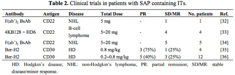
The table below[2] highlights antibody-saporin conjugates approved by the FDA for Phase I/II clinical trials in humans. The therapeutics listed below were administered intravenously and imply what the FDA accepted as non-toxic levels of saporin-based conjugates in these studies.
Looking more closely at the study by French et al.,[3] several milligrams of antibody conjugate were repeatedly injected into human patients under a FDA regulated clinical trial and peak serum levels tested, demonstrating rapid clearing of saporin from the system.
As a company that specializes in Saporin, our two-plus decades of experience working with the protein in research, preclinical, and clinical environments has taught us that with minimal standard laboratory precautions users are not at any real risk of toxic effects. Even our CSO, after 30+ years of working with Saporin exhibits undetectable levels of Saporin antibodies in his blood!
Vierck C, Yezierski R, Wiley R (2016) Pain sensitivity following loss of cholinergic basal forebrain (CBF) neurons in the rat. Neuroscience 319:23-34. doi: 10.1016/j.neuroscience.2016.01.038
Objective: There is a large amount of research on the involvement of cholinergic mechanisms on spinal transmission of pain signals, indicating that cholinergic agonists can attenuate this kind of pain. In contrast, some studies have shown affective reactions to pain are suppressed by cholinergic antagonists. The authors investigated the disagreement between reflexive and affective reactions.
Summary: Lesioned rats displayed decreased escape from thermal stimulation, as well as loss of the normal hyperalgesic effect of sound stress. Results indicate that the basal forebrain cholinergic system plays a role in central processing of pain.
Usage: Administration of 192-IgG-SAP with a 4-μg injection into the left lateral ventricle of rats. Animals were tested in temperature escape and sound stress models.
Schwartz M, Nguyen A, Warrier D, Palmerston J, Thomas A, Morairty S, Neylan T, Kilduff T (2016) Locus coeruleus and tuberomammillary nuclei ablations attenuate hypocretin/orexin antagonist-mediated rem sleep. eNeuro 3:ENEURO.0018-0016.2016. doi: 10.1523/ENEURO.0018-16.2016
Summary: To examine the mechanism by which the Orexin 1r/Orexin 2r antagonist almorexant decreases wakefulness and increases NREM and REM sleep the authors utilized Anti-DBH-SAP (Cat. #IT-03) and Orexin-B-SAP (Cat. #IT-20). Rats received 3-μg injections of Anti-DBH-SAP into the LC, or bilateral 57-80 ng injections of Orexin-SAP into the TMN. Both conjugates attenuated the increased REM sleep seen upon administration of almorexant without altering almorexant-induced changes in NREM sleep.