Roland JJ, Stewart AL, Janke KL, Gielow MR, Kostek JA, Savage LM, Servatius RJ, Pang KC (2014) Medial septum-diagonal band of Broca (MSDB) GABAergic regulation of hippocampal acetylcholine efflux is dependent on cognitive demands. J Neurosci 34(2):506-514. doi: 10.1523/JNEUROSCI.2352-13.2014
Summary: GABAergic and cholinergic neurons in the medial septum-diagonal band of Broca (MSDB) are both involved with spatial memory. In order to better understand the relationship between these two neuronal populations the authors administered 552.5 ng of GAT-1-SAP (Cat. #IT-32) to the MSDB of rats in several injections. Using a combination of behavioral assays and in vivo microdialysis it was shown that GAT-1-SAP lesions impaired hippocampal acetylcholine efflux as well as performance in the non-matching to position with delay test. The data indicate that GABAergic MSDB neurons are important during high memory load conditions.
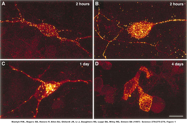
Q: How long does it take to see the cell death occurring from the use of targeted toxins using saporin? Is there a time course of hours or days?
A: The figure below illustrates the time course of cell death very effectively. Internalization and cytotoxicity of SP-SAP in primary cultures of neonatal spinal cord neurons. Confocal image of neurons where the Substance P receptor; NK1R (SPR) immunofluorescence (A, C, D) appears red, areas of concentrated SPR immunofluorescence appear yellow. (A, C, and D) SPR immunofluorescence in neurons 2 hours, 1 day, and 4 days, respectively, after treatment with SP-SAP. (B) Confocal image showing SAP immunofluorescence (yellow) 2 hours after SP-SAP treatment.
These images were projected from 14 optical sections acquired at 0.8-mm intervals with a 603 lens. Bar, 25 mm.
It is recommended that you wait for two weeks to allow for all debris to be cleared and the animal to regain normal eating and sleeping habits.
Weisshaar CL, Winkelstein BA (2014) Ablating spinal NK1-bearing neurons eliminates the development of pain and reduces spinal neuronal hyperexcitability and inflammation from mechanical joint injury in the rat. J Pain 15(4):378-386. doi: 10.1016/j.jpain.2013.12.003
Summary: A high percentage of chronic neck pain involves the facet joint. Although the facet joint is innvervated by peptide-responsive nociceptive afferents, the role of these cells in the development and modulation of nociceptive signaling remains unclear. Using a previously developed rat model of facet joint injury, the authors examined the role of neurokinin-1 receptor-expressing spinal cells in this pathway. Rats received 100 ng SSP-SAP (Cat. #IT-11) via lumbar puncture. Blank-SAP (Cat. #IT-21) was used as a control. The results demonstrate that spinal NK1r-expressing cells are essential for nociception and inflammation due to a mechanical joint injury.
Takakura AC, Barna BF, Cruz JC, Colombari E, Moreira TS (2014) Phox2b-expressing retrotrapezoid neurons and the integration of central and peripheral chemosensory control of breathing in conscious rats. Exp Physiol 99(3):571-585. doi: 10.1113/expphysiol.2013.076752
Summary: Previous work has shown that lesions to the retrotrapezoid nucleus (RTN) have at least a modest effect on breathing, but it is unclear whether those lesions affected the entire nucleus or were incomplete. The authors used bilateral lesions of the RTN with 0.3 to 1.2 ng total of SSP-SAP (Cat. #IT-11) to eliminate neurokinin-1 receptor-expressing neurons; these are also Phox2b+TH- neurons. The results indicate that loss of Phox2b(+)TH(-) neurons may cause deficits seen after RTN lesion, and help define the ways in which these cells are involved in controlling central and peripheral chemoreflexes.
Freiria-Oliveira AH, Blanch GT, De Paula PM, Menani JV, Colombari DS (2013) Lesion of the commissural nucleus of the solitary tract/A2 noradrenergic neurons facilitates the activation of angiotensinergic mechanisms in response to hemorrhage. Neuroscience 254:196-204. doi: 10.1016/j.neuroscience.2013.09.017
Summary: Previous work has generated conflicting data on the role of catecholaminergic A2 neurons in the nucleus of the solitary tract (NTS) in control of arterial pressure lability. The authors used Anti-DBH-SAP (Cat. #IT-03) to lesion these neurons in a hypotensive hemorrhage model. Rats received two injections of 12.6 ng into the commissural NTS. Mouse IgG-SAP (Cat. #IT-18) was used as a control. The lesioned animals quickly recovered from hypotension, but were impaired by the icv administration of losartan.
Summary: Although mainly known for their involvement in the control of arterial pressure, C1 neurons are also suspected to participate in numerous other physiological processes such as neuroendocrine response, glucose homeostasis, food consumption, and others. This review discusses the role of these neurons as ’emergency medical technicians’ – cells that produce and modulate physiological survival responses to acute physical stress. The use of Anti-DBH-SAP (Cat. #IT-03) to delineate C1 neurons in the rostral ventrolateral aspect of the medulla oblongata is discussed.
Grupe M, Paolone G, Jensen AA, Sandager-Nielsen K, Sarter M, Grunnet M (2013) Selective potentiation of (alpha4)3(beta2)2 nicotinic acetylcholine receptors augments amplitudes of prefrontal acetylcholine- and nicotine-evoked glutamatergic transients in rats. Biochem Pharmacol 86(10):1487-1496. doi: 10.1016/j.bcp.2013.09.005
Summary: Nicotinic acetylcholine receptors (nAChR) are involved in a wide range of processes in the central nervous system, many having to do with higher cognitive functions. In order to better understand how these receptors mediate attentional performance, the authors investigated glutamate release under varying conditions. In one series of experiments rats received a 160-ng injection of 192-IgG-SAP (Cat. #IT-01) into the right medial prefrontal cortex. The resulting decrease in glutamate release after the cholinergic lesion adds to the data indicating that positive modulation of nAChR may help alleviate attentional impairments caused by some brain disorders.
Romano A, Potes CS, Tempesta B, Cassano T, Cuomo V, Lutz T, Gaetani S (2013) Hindbrain noradrenergic input to the hypothalamic PVN mediates the activation of oxytocinergic neurons induced by the satiety factor oleoylethanolamide. Am J Physiol Endocrinol Metab 305(10):E1266-73. doi: 10.1152/ajpendo.00411.2013
Summary: Feeding behavior and energy balance are in part controlled by signals from the gut. Oleoylethanolamide (OEA) is an acylethanolamide that is thought to play a role in this network. Since peripheral administration of OEA has effects on the nucleus of the solitary tract (NTS) and paraventricular nucleus (PVN) the authors investigated the role of noradrenergic afferent input to these areas. Rats received bilateral 84-ng injections of Anti-DBH-SAP (Cat. #IT-03) into the PVN. Mouse IgG-SAP (Cat. #IT-18) was used as a control.
Zajo KN, Fadel JR, Burk JA (2013) Assessment of the contributions of baseline performance and prefrontal cortical cholinergic projections to orexin A-induced attentional enhancement. Neuroscience 2013 Abstracts 854.02. Society for Neuroscience, San Diego, CA.
Summary: Orexinergic neurons innervate several brain regions including the basal forebrain, a structure known to be crucial for normal attentional performance in rats. Our previous research demonstrated that orexin receptor blockade impairs attention and that infusions of orexin A into the lateral ventricle enhance attentional performance in animals that have just reached criteria for stable performance levels on a sustained attention task. Our current research investigated whether more highly trained animals show orexin A-induced enhancement of attentional performance and whether basal forebrain cholinergic inputs to the medial prefrontal cortex were necessary for orexin A-induced attentional enhancement. Male FBNF1 hybrid rats were trained in a sustained attention task that required discrimination of visual signals (500, 100 or 25-ms illumination of a central panel light) from trials when no signal was presented. After stable performance levels were established, rats received both intraventricular guide cannula implantation and infusions of either the immunotoxin 192IgG-saporin or vehicle into the medial prefrontal cortex. Postsurgically, rats were retrained to stable performance levels and then received infusions of 0 (vehicle), 10, 100 or 1000pM orexin A in a counterbalanced order prior to task performance. On infusion days, rats were exposed to a version of the task which increased attentional demands by presenting a visual distracter during the middle block of trials within a testing session. In rats trained to higher performance levels, intraventricular orexin A infusions did not significantly enhance attentional performance. Loss of cholinergic projections to the medial prefrontal cortex decreased attentional performance, particularly when a visual distracter was presented. Attentional performance was unaffected in lesioned rats when orexin A was infused into the lateral ventricle. Our findings suggest that orexin A-induced attentional enhancement may be dependent upon baseline performance levels and possibly the integrity of the basal forebrain cholinergic projections to the medial prefrontal cortex.
Khan D, Owens E, Zaben M, Dunnett SB, Gray WP (2013) CD4+ T lymphocytes interact with microglia to modulate hippocampal neurogenesis. Neuroscience 2013 Abstracts 699.04. Society for Neuroscience, San Diego, CA.
Summary: Hippocampal neurogenesis occurs within the subgranular zone of the dentate gyrus and is important for learning and memory. Neurogenesis is impaired in patients with chronic temporal lobe epilepsy, an observation that may account for the learning and memory deficits that these patients commonly have. Emerging literature demonstrates that CD4+ T lymphocytes increase neurogenesis and enhance cognition; however, the exact mechanisms remain undetermined. Vasoactive Intestinal Peptide (VIP) receptors are expressed on T lymphocytes, microglia and hippocampal progenitor cells, hence this study was designed to investigate VIP’s role in mediating neuro-immune modulation. Hippocampal cultures (P7-10 Sprague Dawley rats) were generated and maintained for 3 days in vitro (DIV) and treated with 5% supernatant generated from C57/Bl6 mouse spleen using a CD4+ T lymphocyte isolation kit. BrdU and experimental conditions were added for the terminal 6 hours before fixation and then processed for BrdU and nestin. For phenotype analysis, experimental conditions were added at 3DIV and fixed at 6DIV to be processed for nestin and TuJ1. To deplete microglia, Mac-1-SAP was added at 2DIV for 24 hours before experimental conditions were added. 5% T lymphocytes supernatant increased proliferation of hippocampal nestin-expressing cells; an effect that is further enhanced under VIP treatment via VPAC1 receptor subtype. Examining potential cytokine mediators of this effect, PCR analysis showed 6-fold increase in IL-4 mRNA expression, and IL-4 antagonist abolished VIP proliferative effects. Using Mac-1-SAP to account for microglial involvement by depleting microglia, VIP proliferative effects were abolished. Our phenotyping studies also demonstrated an additional neurogenic effect under VIP treated supernatant compared to standard control conditions. Taken together, these results show VPAC1 receptor subtype expressed by CD4+ T lymphocytes mediates VIP proliferative effects on hippocampal cells via IL-4 cytokine release. Microglia mediates VIP proliferative effects. While we demonstrated before that VPAC2 mediates hippocampal progenitor cell survival, the findings of this study strongly implicate VPAC1 receptor as a neuro-immune mediator of hippocampal neurogenesis, and from a therapeutic perspective, shows that the effect can be pharmacologically manipulated.