- Today’s topic is time-course for our saporin conjugates. Common questions we get are (1) how long does it takes to see cell death, (2) how long should I wait before performing histology on an animal, or (3) how long before I see behavioral changes in an animal?
- In this image from Mantyh et al. (1997), we are looking at confocal imagery of the binding and internalization of our peptide conjugate SP-SAP to the NK1r receptor in primary cultures of neonatal spinal cord neurons.
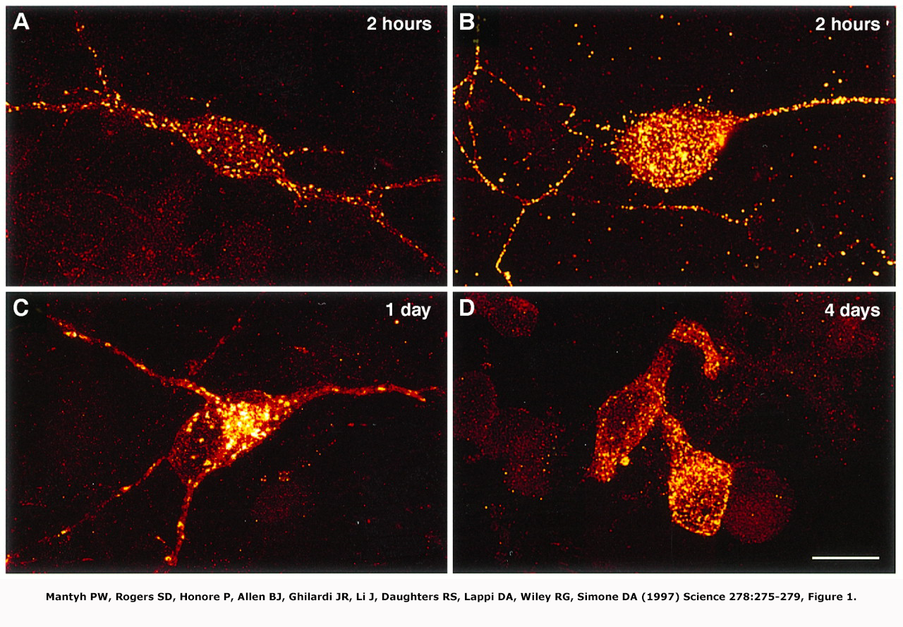
- As you can see, conjugate binding occurs immediately where within hours, the SP-SAP has recognized and bound to the NK1 receptor. Here we see the areas of concentrated NK1R expression marked by yellow immunofluorescence.
- But how long until you see cell death? Here is a cytotoxicity graph of in vitro data of our antibody conjugate 192-IgG-SAP. These are typical data after cells have been treated for 3 days, which is standard protocol.
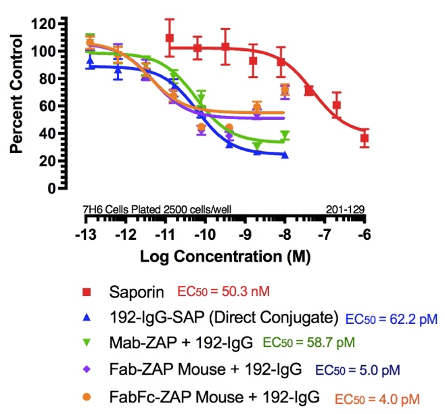
- Waite et al. (1994) used 192-IgG-SAP and showed the appearance of behavioral effects associated with neuronal loss at day four and plateauing at day 7.
- This coincides with the time course seen in vitro. At this point, microglia will infiltrate, but stop at 7 days, which is probably the peak day for infiltration. Once there is complete removal of the detritus, microglia down-regulate and at 14 days all debris is cleared and the animal begins to regain normal eating and sleeping habits. This is the idea behind waiting about 2 weeks before performing histology.
episode27, episode49