Contributed by Ai-Jun Li, Qing Wang, Thu T. Dinh and Sue Ritter
Programs in Neuroscience, Washington State University, Pullman, Washington 99164-6520, USA
Leptin is a fat tissue-derived hormone with widespread actions in brain and peripheral tissues. Leptin’s actions are mediated by the long isoform of the leptin receptor, OB-Rb, which has a lengthy intracellular region containing several motifs required for signal transduction via the JAK2/STAT3 pathway. Leptin profoundly influences food intake and body weight, in large part by its actions on OB- Rb receptors in the arcuate nucleus (Arc) in the basomedial hypothalamus. Two populations of neurons in the Arc that are importantly involved in these functions are neuropeptide Y (NPY) and agouti gene-related protein (AGRP) co-expressing neurons and pro-opiomelanocortin (POMC) and cocaine- and amphetamine-regulated transcript (CART) co-expressing neurons. The majority of both NPY/AGRP and POMC/CART neurons are OB-Rb-positive. Exogenous administration of leptin excites and increases phospho-STAT3 expression in POMC/CART neurons and inhibits NPY neurons in the Arc.
In the present study, we used a novel leptin-saporin conjugate (Lep-SAP, Cat. #IT-47), a targeted toxin developed recently by Advanced Targeting Systems, to lesion OB-Rb-expressing NPY/AGRP and POMC/CART neurons in Arc. Binding of the Lep-SAP to the OB-Rb receptor is the mechanism for selective internalization of saporin, a ribosomal toxin that destroys the cell. The goals of our study were (1) to determine whether this new Lep-SAP conjugate is an effective and selective lesioning agent and (2) to use this targeted toxin to further examine the role played by Arc OB-Rb-expressing neurons in the actions of endogenous leptin.
A dose-response analysis was conducted to determine the dose, volume and injection sites for lesioning the Arc. One or two sites per side were injected. Lesions were analyzed by quantification of NPY/AGRP and POMC neurons in the Arc by immunohistochemistry (IHC) and real-time PCR. Results showed that bilateral injections of 57 ng in 50 nl per injection site at two sites in the sagittal plane produced a lesion that was confined to, but extended throughout, the Arc.
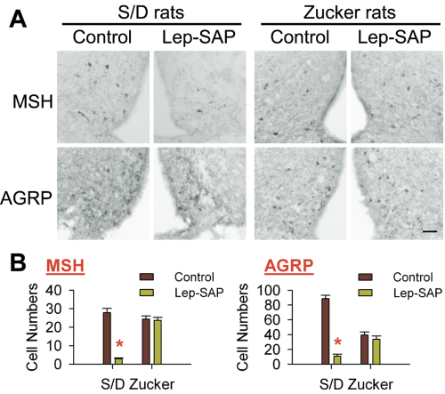
To evaluate the specificity of Lep-SAP for lesioning leptin receptor-expressing neurons in vivo, unilateral injections of Lep-SAP or control Blank-SAP (B-SAP, Cat. #IT-21) were made into the Arc in Sprague Dawley (S/D) rats and Zucker fa/fa fatty rats, which have a mutation on the extracellular domain of the leptin receptor. IHC staining was performed to detect hypothalamic neurons positive for AGRP and the POMC- derived peptide, α-melanocyte-stimulating hormone (α-MSH). Figure 1 shows that in S/D rats, numbers of α-MSH- and AGRP-positive cells were significantly decreased on the Lep-SAP injected side to 16-18% of the numbers found on the non- injected side. In contrast, in Zucker fa/fa rats, numbers of α-MSH and AGRP neurons did not differ on the injected versus the noninjected sides. The effective reduction of leptin- receptor expressing neurons in the Arc of S/D rats by Lep-SAP and the failure of the toxin to lesion these neurons in the Zucker fa/fa rats lacking a functional leptin receptor indicates that Lep-SAP is an effective lesioning agent and that its internalization is dependent on the Ob-Rb receptor.
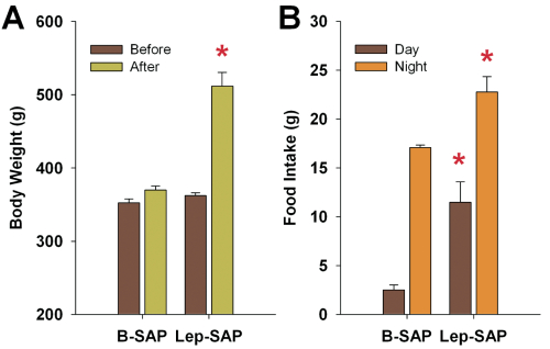
To evaluate the effect of Arc Lep-SAP on food intake and body weight, Lep-SAP or B-SAP control was injected bilaterally into Arc. We found that Lep-SAP rats rapidly became hyperphagic and obese after the injection and maintained a level of intake that was 2-fold greater than that of the B-SAP rats (Fig. 2). Diurnal rhythms of food intake were also altered. Daytime feeding was significantly enhanced. Lep-SAP rats did not respond to central leptin administration, had elevated plasma levels of glucose, free fatty acids, triglycerides, insulin, and leptin, and had excessive fat deposits in liver, and in white and brown fat pads. Expression of clock-related genes was measured at a single time point in the light period by real-time PCR. Bmal1 expression in whole hypothalamus, liver, and white fat tissue of Lep-SAP rats was significantly decreased. Per1 expression was increased in liver and decreased in white fat tissue, compared to B-SAP controls. Per1 expression in the hypothalamus did not differ between Lep-SAP and B-SAP rats.
These results show that Lep-SAP effectively lesioned Arc NPY/AGRP and POMC neurons in a leptin receptor-dependent manner. In addition, these results support previous findings showing that Arc NPY/AGRP and POMC neurons play a critical role in the control of food intake and energy expenditure. Finally, we report new findings indicating a role for the Arc in the diurnal patterning of food intake and in central and peripheral clock-related gene expression. Thus, Lep-SAP is a useful new tool for studying the functions of leptin receptor-expressing neurons in specific brain sites.
Reference: (back to top)
- A.-J. LI, Q. WANG, T. T. DINH, S. RITTER, Leptin-saporin injection into the arcuate nucleus lesions NPY/AGRP and POMC neurons and produces hyperphagia, obesity and changes in diurnal feeding patterns in rats. Program No 374.5. 2009 Neuroscience Meeting Planner. Chicago, IL: Society for Neuroscience, 2009.