Advanced Targeting Systems announces three new antibodies to G protein-coupled receptors that will be quite helpful for a number of researchers studying somatostatin and substance P.
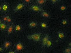
For somatostatin study, we already market excellent rabbit polyclonals to somatostatin-14 (Cat. #AB-04) and somatostatin-28 (Cat. #AB-05). We now announce a new mouse monoclonal antibody to somatostatin receptor 1 (SSTr1, Cat. #AB-N35). Figure 1 shows staining of fixed cells and a distribution of staining typical for a membrane-bound protein. This protein’s antigen is to an extracellular domain of the rat sequence and it is an IgM.
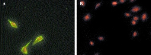
We are also releasing a mouse monoclonal to SSTr5 (Cat. #AB-N24). Figure 2 shows staining with this IgM in panel A and normal mouse IgM in panel B, demonstrating specificity. It is also made to an extracellular sequence of the protein.
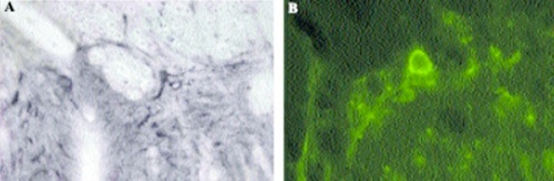
We have created a rabbit polyclonal to a sequence from the dog substance P (NK-1) receptor (Cat. #AB-N33AP). Photomicrographs of the use of this antibody are seen in Figure 3. As researchers in this field, we know the difficulty of finding a good, consistent source of this antibody; sequence homologies are very close with rat and human (one amino acid change out of 15), so this antibody should be excellent in other species as well. The antigen for this antibody is a peptide near the C- terminus.