Contributed by Dr. Naoki Yoshimura, Dept Urology/Pharmacology, Univ Pittsburgh School of Medicine, Pittsburgh PA 15213.
Dr. Yoshimura summarizes his work with IB4-SAP. A complete report was published in Eur J Neurosci 20(2):474-482, 2004.
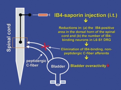
It has been demonstrated that hyperexcitability of C-fibers in bladder afferent pathways can contribute to bladder overactivity and/or bladder pain under pathological conditions such as spinal cord injury or chronic cystitis. We have also previously demonstrated that peptidergic and IB4-binding non-peptidergic C-fiber populations are present in the afferent pathways to the bladder and that the relative proportion of these two populations of neurons is different from that in the somatic afferents pathways. Therefore, in this present study, we investigated the effects of intrathecal application of the IB4-saporin (IB4-SAP, Cat. #IT-10) conjugate at the level of L6-S1 spinal cord, where bladder afferent pathways terminate, on the normal bladder function and bladder overactivity induced by bladder irritation. The goal of this study was to elucidate the functional role of IB4-binding afferent pathways in bladder function.
We have found that intrathecal treatment with IB4-saporin at the level of L6-S1 spinal cord reduced IB4 afferent nerve terminal staining in the lamina II of the dorsal horn of L6 spinal cord as well as the number of IB4-binding neurons in L6 DRG (Fig. 1 and Fig. 2). IB4-saporin suppressed bladder overactivity induced by intravesical capsaicin or ATP without affecting normal micturition. These results indicate that elimination of IB4-binding afferents by IB4-SAP is effective for the treatment of bladder overactivity induced by bladder irritation. Thus, targeting IB4-binding, presumed non-peptidergic afferent pathways, which are sensitive to capsaicin and ATP, may be an effective treatment for overactivity and/or visceral pain responses in the bladder.
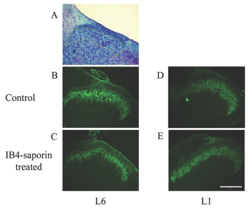
Histochemical staining of the L1 and L6 spinal cord in normal rat (A). IB4-conjugated FITC staining of the L6 spinal cord from control (B) and IB4-saporin-treated rats (C) 3 weeks after the treatment. IB4-conjugated FITC staining of the L1 spinal cord from control (D) and IB4-saporin-treated rats (E) 3 weeks after the treatment.
The lamina II area identified by Nissl’s staining is indicated by dashed lines in A. Note that the staining density of IB4-binding afferent nerve terminals in the lamina II of the L6 spinal cord was depleted after the IB4-saporin treatment. Scale bar, 200 µm.
Printed with permission of Blackwell Publishing
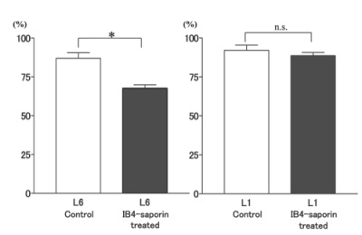
The area positively stained with IB4 in the lamina II of the dorsal horn in the L1 and L6 spinal cord from control and IB4-saporin-treated rats (3 weeks after the treatment). Data are expressed as percentages of the positively stained area in total lamina II area (100%). *P < 0.01 (unpaired t-test)
Printed with permission of Blackwell Publishing