Contributed by Patrick Shramm, Product Manager for Fab-pHast
We are pleased to announce a new set of tools to rapidly screen antibodies, Fab-pHast conjugates. The ATS pHAST product line provides the FASTEST results in quantitative testing of your antibody’s specific-binding and internalization. Fab-pHast contains a pH-dependent fluorescent reporter that increases intensity in acidic surroundings, such as the environment inside a cell. You just mix the Fab-pHast with your antibody, add to cells, and in less than 24 hours you have illuminated your lead antibody candidates!
pHast Ab Internalization Assay: Parental HEK-293 cells, and HEK-293 cells transfected with the p75 receptor, were plated in a 96-well plate overnight. Titrated 192-IgG antibody (Cat. AB-N43) was incubated at RT with 50 nM of Fab-pHast Mouse (Cat. PH-02) for 20 minutes prior to addition to cells. Plates were incubated overnight to allow maximum internalization, but a few hours is sufficient for detection. Plates were read on a Spectra Max Gemini EM (Ex: 532nm/Em: 560nm). Data analysis was done by PRISM (GraphPad, San Diego).
When ordering, be sure to choose the appropriate species for your primary antibody. These products are designed to provide an EC50 by way of a fluorescence-detecting plate reader. Your best candidates reach maximum intensity overnight, but you can make quantitative decisions in just a few hours. Other applications include qualitative visualization under a fluorescent microscope or analysis via flow cytometry.
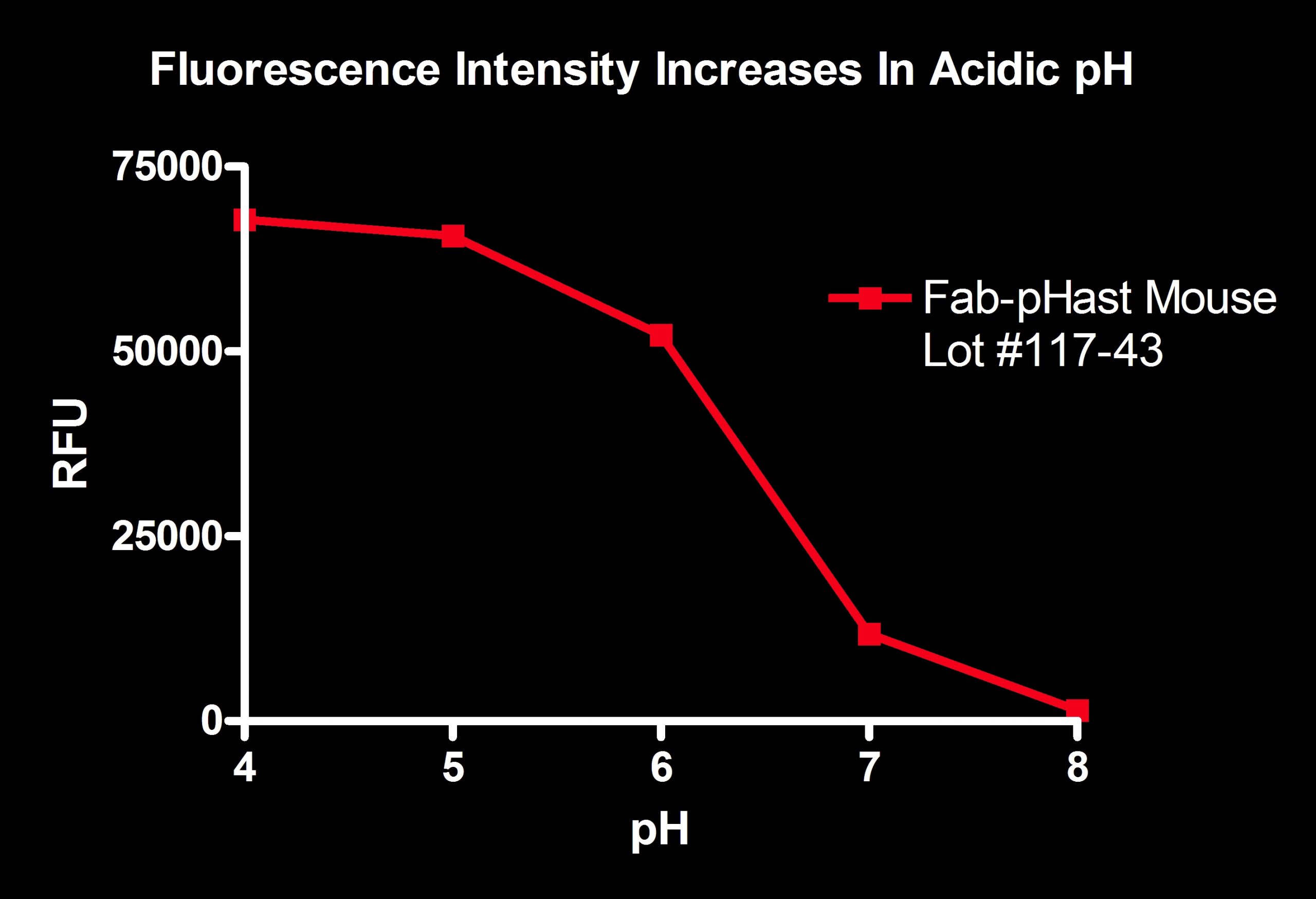
Fluorescence (RFU) is shown as a function of pH for Fab-pHast Mouse (Cat. PH-02). The more acidic pH shows a large amount of fluorescence, while the basic pH shows almost no fluorescence. Plates read on a Spectra Max Gemini EM (Ex: 532nm/Em: 560nm).
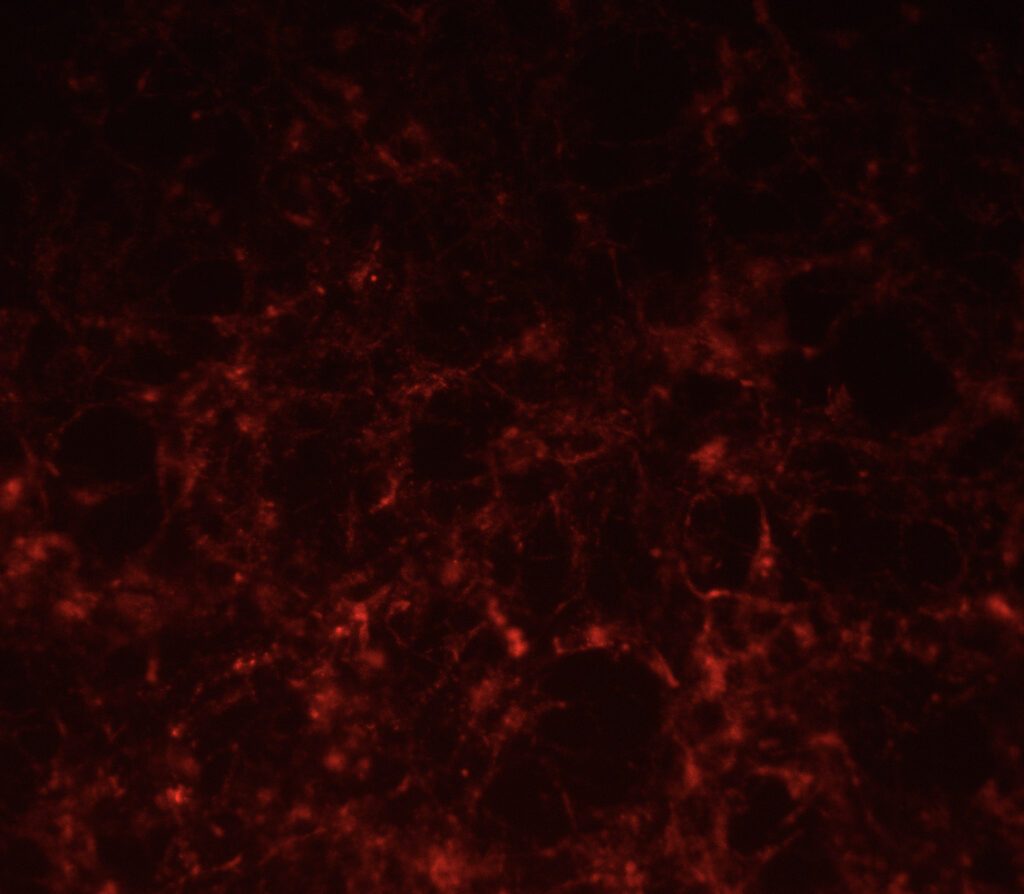
ICC image of Anti-NGFr (192-IgG, Cat. AB-N43) illuminated with Fab-pHast Mouse (Cat. PH-02). HEK-293 cells, transfected with the p75 receptor, were plated at 20,000 cells/well in a 96-well plate and allowed to adhere overnight. 10 nM of the primary antibody was incubated at RT with 30 nM of Fab-pHast. Cells were incubated overnight to allow maximum internalization, although internalization can be detected in a few hours. Cells were analyzed on a Leica microscope under 20X magnification using a Y3 filter cube.